Quality is Top Priority
Ultrasound, which is sound above the human hearing limit of approximately 16 kHz, is applied in numerous ways in medical engineering and research. Applications range from distance measurement and object recognition, filling level or flow rate measurements, through high-resolution material tests, up to medical diagnosis and therapy. Here, piezo elements often play a key role, as is also the case in a new system for the wireless monitoring of mother and child during pregnancy and birth (Image 1).

The creation and detection of ultrasound, for example, is a classic piezo application because the piezo element starts to oscillate when an a.c. voltage is applied. An electric voltage is generated under force (direct piezo effect) and the dimensions change under the influence of an electric field (inverse piezo effect). In other words, piezo elements convert mechanical into electrical energy and vice versa, and are therefore referred to as ultrasound transducers. The piezo effect is based exclusively on displacements within the crystal lattice of the piezoelectric element. There is therefore no mechanical friction and no wear in the classical sense, and sensitivity is high. Even the smallest deformation results in immediately measurable charge transfer. Inversely, a small electrical voltage results in immediate displacement. Piezo elements can therefore be used as sensors and actuators at the same time, they not only generate ultrasound but also detect it. The short response times and the high dynamics of their displacement naturally also benefit ultrasound generation.
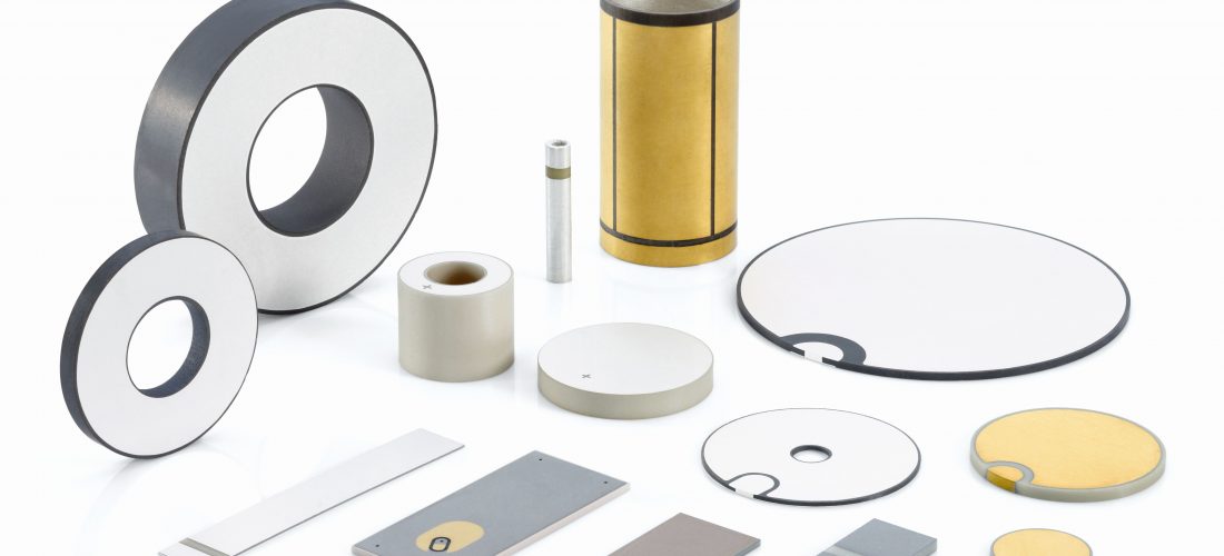
In addition, piezo elements can also be easily adapted to the respective application requirements as different geometric variants and resonance frequencies can also be realized (Image 2) in addition to the material selection specific to each application. However, depending on the application, high quality standards need to be met when manufacturing the piezos. In the case of ultrasound transducers for cardiotocographs (fetal monitors or CTGs), i.e. the monitoring of the heart rate of the unborn child, these requirements are extremely high.
Fetal Monitoring: Identical Wireless Monitoring Even for Triplets
Philips Medizin Systeme Böblingen GmbH, Germany, is regarded as the market leader in patient monitoring systems. Now the successful company has developed a new wireless operating cardiotocograph (see Image 1). With the aid of this machine, all fetal heart rates can be recorded in sync, i.e. with the same impulse density, even for triplet pregnancies. All unborn children therefore receive the same monitoring quality, they are each monitored at a repetition rate of 3000 ultrasound bursts per second.
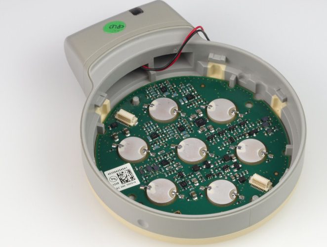
Image 3 shows the interior of the ultrasound transducer. Seven identical piezo elements are arranged symmetrically and operate both as transmitter and receiver for ultrasound. The received signal is separated from the carrier in a signal processing stage, filtered and emitted acoustically to facilitate the positioning of the transducer on the one hand, and on the other fed to a further signal processing stage. There the signal is rectified, filtered and reduced in frequency. With the aid of autocorrelation, the fetal heart frequency is calculated from this signal, ideally for every heart beat. The basis for these elaborate calculations is provided by the so-called Doppler effect, i.e. the frequency shift between an emitted and received, reflected wave front of ultrasound waves on a particular transducer is evaluated. Reflection is, for example, caused by moving structures such as the heart muscle of the unborn child. The fetal heart rate can thus be detected and monitored.
Quality Assurance: Optimum Utrasound Parameters, Surface Cleanliness and Bonding
Stringent quality requirements are applied to the selection of suitable piezo elements. Only if the piezo elements meet the required criteria, can the control of the transducer and the evaluation algorithms be matched in an optimum manner. Only this will result in the precise results which make seamless monitoring possible even for triplets. Important prerequisites therefore include consistent ultrasound performance of the piezo elements as transmitters and receivers, as well as, for example, bonding quality. The adhesive joint needs to be as strong as possible on the one hand to conduct the ultrasound signals optimally to the surface and couple them to the evaluating electronics, and on the other it should remain elastic to absorb impacts. In addition, the transducer must be robust enough so as not to be damaged if dropped accidentally during daily clinical routine.
In the end, the disc-shaped piezo elements manufactured by PI Ceramic GmbH, which belongs to the PI Group proved to be ideal for the application. The piezos “Made in Germany” were convincing due to their narrow tolerances of piezo-electric parameters relevant for ultrasound performance, such as resonance frequency, electrical capacity and coupling coefficient. In addition, the piezo elements from PI Ceramic displayed an extremely high surface cleanliness. This allows optimum, solid and yet elastic bonding. When dropped from a height of 1.5 m, the worst that can happen to the transducer is cosmetic damage.
Homogeneous Ultrasound Performance
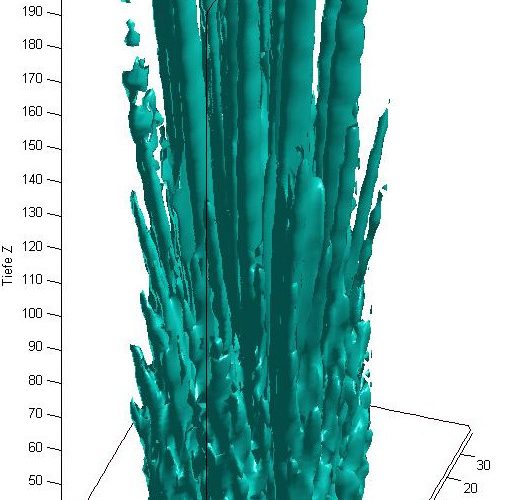
During the manufacturing process, the quality of the adhesive joints is monitored continuously by appropriate measurement of the bonding force. And, of course, the signal strength is also checked intensively, to ensure that all seven piezo elements in the finished transducer really operate at equal signal strengths and also react in the same manner when receiving signals. The acoustic field is measured spatially using a three-axis measuring system to optimize the sound field. The objective is to obtain an even, ideally cylinder-shaped sound field.
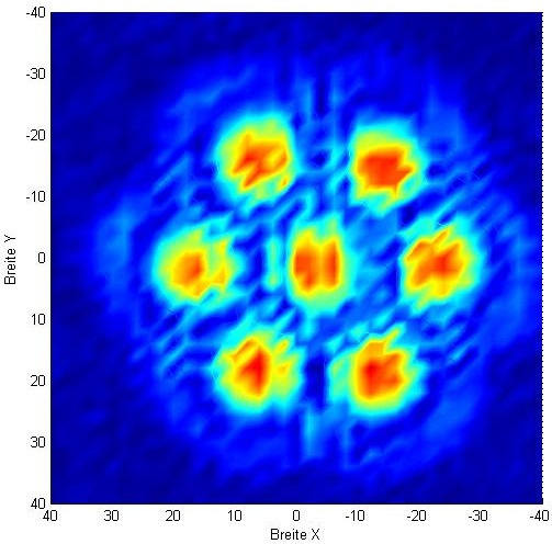
Images 4 and 5 illustrate a horizontal cross-section through the sound field as well as a three-dimensional representation of the sound intensity as recorded in a water tank. The result of the quality-oriented manufacturing process is a largely homogeneous ultrasound performance of the transducer. In addition, this also allows to guarantee adequate ultrasound penetration at all times. The latter is important as the trend towards obesity is continuing in nearly all countries, so that increasingly more tissue needs to be penetrated by the ultrasound. At the same time this ensures that the energy transfer into the tissue is kept as low as possible. It lies considerably below the values specified in the relevant standards.
Manufacturing of the piezo elements was also subject to further application-specific requirements. For example, the polarity markings of the piezo elements had to be applied according to the user specifications in such a way that they could not be confused during cleaning which is obligatory prior to assembly in the transducer. Handling at the user’s location has been simplified considerably through the well visible polarity markings. The piezo elements “Made in Germany” proved convincing for the new wireless fetal monitoring system, not only from a technical point of view, but also in terms of practicality.