Define hyphae
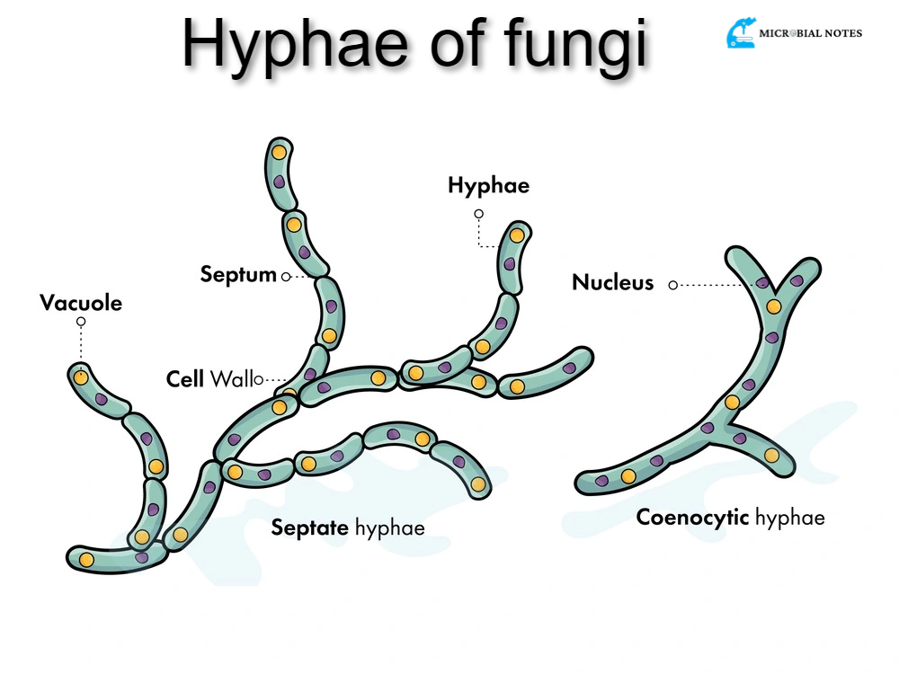
Hyphae are the long filament branches that are seen in fungi and actinobacteria (shown below). Mycelium is the aggregate name for these species’ hyphae, which are important structures required for growth.
Explain the structure of hyphae
Each hypha contains internal septa that divide the cells and at least one cell is enclosed by a protective cell wall, often formed of chitin. Septa play a crucial role in cellular communication by allowing wide apertures for the passage of cellular organelles like ribosomes. However, not all fungus species have septa. Hyphae typically range in size from 4 to 6 microns.
Explain the growth of hyphae.
By expanding the cell walls and internal components from the tips, hyphae proliferate. By storing vesicles from the Golgi apparatus and releasing them along the apex of the hypha, an organelle known as the spitzenkorper helps the development of new cell walls and membrane structures during tip growth.
The release of the vesicle contents, which form the cell wall, and the vesicle membranes, which produce a new cell membrane, causes the tip of the hypha to extend as the spitzenkorper moves. New septa can develop as the hypha grows to further split the cells inside. The creation of a new tip from a hypha or the division of a developing tip causes the characteristic branching of hyphae.
The function of hyphae
Depending on the particular needs of each fungus species, hyphae serve a variety of distinct functions. Here is a list of some of the most well-known hyphae functions:
Nutrient absorption from the host
Some parasitic fungal hyphae are tailored for nutrient absorption inside a particular host. These hyphae have specialized ends known as haustoria that can pierce plant cell walls or other tissues to access nutrients.
Nutrient absorption from soil
Some fungi, like mycorrhizae, have cultivated symbiotic bonds with various vascular plant species. To take nutrients and water from the soil, the fungal creates specialized hyphae called arbuscules, which are located in the roots or phylum of vascular plants. By improving the plant’s ability to obtain nutrients in the soil and promoting its growth, the hyphae help the plant.
Trapping structures
Some fungal species have developed hyphae that are specialized worm-trapping structures, encasing nematode species in nets and rings.
Nutrient transportation
Mycelial chords, which are found in the hyphae of some fungal species, are employed by fungi (such as lichens and mushrooms) to carry nutrients across long distances.
Explain hyphae classification.
Hyphae can be categorized generally according to the following characteristics;
Hyphae Characteristics
An essential tool for distinguishing different fungi species is the hyphae. There are three main traits of hyphae:
Binding
Binding hyphae are heavily branching and have a thick cell wall.
Generic
Generic hyphae are often less differentiated and contain a thin cell wall and many septa. Additionally, generative hyphae can produce structures required for reproduction when they are embedded in other substances, such as gelatin or mucilage. Generative hyphae are normally present in all fungi species.
Skeletal
The cell wall of skeletal hyphae is lengthy and thick, and it has few septa. Skeletal hyphae may also be of the fusiform variety, characterized by large middle and tapered ends.
Hyphae Composition
Based on the hyphal systems they possess, fungus species are further divided into different categories. Four basic subtypes exist:
Monomitic
Although almost all fungi have generative hyphae, this variety of fungi is the only one that displays it (e.g., agaric mushrooms).
Dimitic
A species that has two different types of hyphae in addition to generative hyphae. Dimitic fungi most frequently occur in generative and skeletal combinations.
Trimitic
Species with all three forms of hyphae are said to be traumatic (generative, binding, and skeletal).
Sarcodimitic and sarcotrimitic
These hyphae are fusiform skeletal hyphae connected to generative hyphae, respectively. Fusiform skeletal hyphae, binding hyphae, and generative hyphae are all present in sarcotrimitic animals.
Hyphae Refraction
Gloeoplerous is the name used to describe hyphae that seem oily or granular under a microscope. Additionally, the hyphae of distinct species are categorized using this word.
Cell division
On the basis of the presence of internal septa, hyphae can be categorized (septate versus aseptate species). The distinction between hyphae and species that form pseudohyphae during cell division is also possible. Incomplete cell division, or pseudohyphae, occurs when the dividing cells do not split. These pseudohyphae are produced by many yeast species.
What is the summary of hyphae?
Eukaryotic creatures known as fungi first arrived on land over 450 million years ago. They are heterotrophs and do not have chloroplasts or other photosynthetic pigments like chlorophyll. A significant component of the environment’s decomposition process is fungi. They play a variety of additional crucial roles in ecosystems as parasites and mutualists, though. External enzymes break down nutrients that are then ingested by the fungus’s mycelium, which is made up of filamentous cells referred to as hyphae.
Chitin forms a substantial cell wall around the hyphae. Yeasts are unicellular fungi that reproduce by budding. Molds are an example of fungi that can reproduce asexually and/or sexually. The life cycle and type of sexual structures created by fungi were two of the main factors used to group them into phyla.
References
https://bio.libretexts.org/Bookshelves/Botany/Botany
Hyphae