US8900282B2 - Light therapy apparatus and methods - Google Patents
Light therapy apparatus and methods Download PDFInfo
- Publication number
- US8900282B2 US8900282B2 US13/895,330 US201313895330A US8900282B2 US 8900282 B2 US8900282 B2 US 8900282B2 US 201313895330 A US201313895330 A US 201313895330A US 8900282 B2 US8900282 B2 US 8900282B2
- Authority
- US
- United States
- Prior art keywords
- light
- light source
- therapy apparatus
- patient
- light therapy
- Prior art date
- Legal status (The legal status is an assumption and is not a legal conclusion. Google has not performed a legal analysis and makes no representation as to the accuracy of the status listed.)
- Expired - Fee Related
Links
- 238000001126 phototherapy Methods 0.000 title claims abstract description 97
- 238000000034 method Methods 0.000 title abstract description 8
- 238000011282 treatment Methods 0.000 claims abstract description 71
- 210000000988 bone and bone Anatomy 0.000 claims abstract description 16
- 208000020084 Bone disease Diseases 0.000 claims abstract description 7
- 210000005069 ears Anatomy 0.000 claims abstract description 5
- 210000004872 soft tissue Anatomy 0.000 claims abstract description 5
- 210000001847 jaw Anatomy 0.000 claims description 17
- 239000000463 material Substances 0.000 claims description 16
- 210000003128 head Anatomy 0.000 claims description 11
- 238000011269 treatment regimen Methods 0.000 claims description 11
- 238000001816 cooling Methods 0.000 claims description 10
- 102000000634 Cytochrome c oxidase subunit IV Human genes 0.000 claims description 5
- 108090000365 Cytochrome-c oxidases Proteins 0.000 claims description 5
- 210000002050 maxilla Anatomy 0.000 claims description 3
- 229920003023 plastic Polymers 0.000 claims description 3
- 239000007788 liquid Substances 0.000 claims description 2
- 230000004044 response Effects 0.000 claims description 2
- 238000010521 absorption reaction Methods 0.000 claims 5
- 230000008878 coupling Effects 0.000 claims 2
- 238000010168 coupling process Methods 0.000 claims 2
- 238000005859 coupling reaction Methods 0.000 claims 2
- 239000000758 substrate Substances 0.000 abstract description 11
- 208000029985 osteonecrosis of the jaw Diseases 0.000 abstract description 5
- 230000001764 biostimulatory effect Effects 0.000 abstract 1
- 210000001519 tissue Anatomy 0.000 description 27
- 238000003491 array Methods 0.000 description 15
- 238000002203 pretreatment Methods 0.000 description 7
- 208000029725 Metabolic bone disease Diseases 0.000 description 6
- 230000035876 healing Effects 0.000 description 6
- 210000000214 mouth Anatomy 0.000 description 6
- 230000000638 stimulation Effects 0.000 description 6
- 238000001356 surgical procedure Methods 0.000 description 6
- 208000028755 loss of height Diseases 0.000 description 5
- 238000002560 therapeutic procedure Methods 0.000 description 5
- 230000001133 acceleration Effects 0.000 description 4
- 230000018044 dehydration Effects 0.000 description 4
- 238000006297 dehydration reaction Methods 0.000 description 4
- 241000973495 Odax pullus Species 0.000 description 3
- 230000006872 improvement Effects 0.000 description 3
- 230000007246 mechanism Effects 0.000 description 3
- 230000003287 optical effect Effects 0.000 description 3
- 230000002159 abnormal effect Effects 0.000 description 2
- 239000000853 adhesive Substances 0.000 description 2
- 230000001070 adhesive effect Effects 0.000 description 2
- 239000002390 adhesive tape Substances 0.000 description 2
- 238000004458 analytical method Methods 0.000 description 2
- 230000008901 benefit Effects 0.000 description 2
- 230000004071 biological effect Effects 0.000 description 2
- 230000008859 change Effects 0.000 description 2
- 239000004053 dental implant Substances 0.000 description 2
- 238000013461 design Methods 0.000 description 2
- 230000007717 exclusion Effects 0.000 description 2
- 230000001815 facial effect Effects 0.000 description 2
- 230000001678 irradiating effect Effects 0.000 description 2
- 230000002045 lasting effect Effects 0.000 description 2
- 230000003902 lesion Effects 0.000 description 2
- 229910052751 metal Inorganic materials 0.000 description 2
- 239000002184 metal Substances 0.000 description 2
- 238000010883 osseointegration Methods 0.000 description 2
- 230000003239 periodontal effect Effects 0.000 description 2
- 201000001245 periodontitis Diseases 0.000 description 2
- 230000005855 radiation Effects 0.000 description 2
- 238000011160 research Methods 0.000 description 2
- 230000008685 targeting Effects 0.000 description 2
- 229920002554 vinyl polymer Polymers 0.000 description 2
- -1 vinyl siloxane Chemical class 0.000 description 2
- 235000012431 wafers Nutrition 0.000 description 2
- 206010061274 Malocclusion Diseases 0.000 description 1
- 208000010191 Osteitis Deformans Diseases 0.000 description 1
- 206010031264 Osteonecrosis Diseases 0.000 description 1
- 208000001132 Osteoporosis Diseases 0.000 description 1
- 208000027067 Paget disease of bone Diseases 0.000 description 1
- 206010052428 Wound Diseases 0.000 description 1
- 208000027418 Wounds and injury Diseases 0.000 description 1
- 238000007792 addition Methods 0.000 description 1
- 229910052782 aluminium Inorganic materials 0.000 description 1
- XAGFODPZIPBFFR-UHFFFAOYSA-N aluminium Chemical compound [Al] XAGFODPZIPBFFR-UHFFFAOYSA-N 0.000 description 1
- 230000002238 attenuated effect Effects 0.000 description 1
- 238000005452 bending Methods 0.000 description 1
- 230000009286 beneficial effect Effects 0.000 description 1
- 208000016738 bone Paget disease Diseases 0.000 description 1
- 230000037182 bone density Effects 0.000 description 1
- 230000008468 bone growth Effects 0.000 description 1
- 230000010072 bone remodeling Effects 0.000 description 1
- 230000001427 coherent effect Effects 0.000 description 1
- 238000004891 communication Methods 0.000 description 1
- 230000001010 compromised effect Effects 0.000 description 1
- 239000004020 conductor Substances 0.000 description 1
- 230000001419 dependent effect Effects 0.000 description 1
- 230000003292 diminished effect Effects 0.000 description 1
- 238000005516 engineering process Methods 0.000 description 1
- 210000000887 face Anatomy 0.000 description 1
- 201000010103 fibrous dysplasia Diseases 0.000 description 1
- 238000003780 insertion Methods 0.000 description 1
- 230000037431 insertion Effects 0.000 description 1
- 230000014759 maintenance of location Effects 0.000 description 1
- 230000004048 modification Effects 0.000 description 1
- 238000012986 modification Methods 0.000 description 1
- 238000012544 monitoring process Methods 0.000 description 1
- 230000011164 ossification Effects 0.000 description 1
- 208000002865 osteopetrosis Diseases 0.000 description 1
- 238000009258 post-therapy Methods 0.000 description 1
- 210000003456 pulmonary alveoli Anatomy 0.000 description 1
- 238000007634 remodeling Methods 0.000 description 1
- 239000012858 resilient material Substances 0.000 description 1
- 238000012552 review Methods 0.000 description 1
- 239000004065 semiconductor Substances 0.000 description 1
- 230000001954 sterilising effect Effects 0.000 description 1
- 238000004659 sterilization and disinfection Methods 0.000 description 1
- 230000004936 stimulating effect Effects 0.000 description 1
- 230000009885 systemic effect Effects 0.000 description 1
- 238000012360 testing method Methods 0.000 description 1
- 238000002604 ultrasonography Methods 0.000 description 1
- 230000000007 visual effect Effects 0.000 description 1
Images


Classifications
-
- A—HUMAN NECESSITIES
- A61—MEDICAL OR VETERINARY SCIENCE; HYGIENE
- A61N—ELECTROTHERAPY; MAGNETOTHERAPY; RADIATION THERAPY; ULTRASOUND THERAPY
- A61N5/00—Radiation therapy
- A61N5/06—Radiation therapy using light
- A61N5/0613—Apparatus adapted for a specific treatment
-
- A—HUMAN NECESSITIES
- A61—MEDICAL OR VETERINARY SCIENCE; HYGIENE
- A61N—ELECTROTHERAPY; MAGNETOTHERAPY; RADIATION THERAPY; ULTRASOUND THERAPY
- A61N5/00—Radiation therapy
- A61N5/06—Radiation therapy using light
- A61N2005/0626—Monitoring, verifying, controlling systems and methods
- A61N2005/0629—Sequential activation of light sources
-
- A—HUMAN NECESSITIES
- A61—MEDICAL OR VETERINARY SCIENCE; HYGIENE
- A61N—ELECTROTHERAPY; MAGNETOTHERAPY; RADIATION THERAPY; ULTRASOUND THERAPY
- A61N5/00—Radiation therapy
- A61N5/06—Radiation therapy using light
- A61N2005/0635—Radiation therapy using light characterised by the body area to be irradiated
- A61N2005/0643—Applicators, probes irradiating specific body areas in close proximity
- A61N2005/0645—Applicators worn by the patient
- A61N2005/0647—Applicators worn by the patient the applicator adapted to be worn on the head
-
- A—HUMAN NECESSITIES
- A61—MEDICAL OR VETERINARY SCIENCE; HYGIENE
- A61N—ELECTROTHERAPY; MAGNETOTHERAPY; RADIATION THERAPY; ULTRASOUND THERAPY
- A61N5/00—Radiation therapy
- A61N5/06—Radiation therapy using light
- A61N2005/065—Light sources therefor
- A61N2005/0651—Diodes
- A61N2005/0652—Arrays of diodes
-
- A—HUMAN NECESSITIES
- A61—MEDICAL OR VETERINARY SCIENCE; HYGIENE
- A61N—ELECTROTHERAPY; MAGNETOTHERAPY; RADIATION THERAPY; ULTRASOUND THERAPY
- A61N5/00—Radiation therapy
- A61N5/06—Radiation therapy using light
- A61N2005/0658—Radiation therapy using light characterised by the wavelength of light used
- A61N2005/0659—Radiation therapy using light characterised by the wavelength of light used infrared
-
- A—HUMAN NECESSITIES
- A61—MEDICAL OR VETERINARY SCIENCE; HYGIENE
- A61N—ELECTROTHERAPY; MAGNETOTHERAPY; RADIATION THERAPY; ULTRASOUND THERAPY
- A61N5/00—Radiation therapy
- A61N5/06—Radiation therapy using light
- A61N2005/0658—Radiation therapy using light characterised by the wavelength of light used
- A61N2005/0662—Visible light
Definitions
- This invention relates to light therapy.
- Apparatus and methods according to the invention may be applied to the treatment of bone disorders and the biostimulation of bone and soft tissue.
- Embodiments of the invention provide apparatus for irradiating tissues of the face and jaw with biologically effective doses of light.
- Light therapy involves irradiating tissues with light.
- Light can stimulate a variety of biological activities in cells and tissues that are compromised in function.
- Light therapy treatment is typically administered by a physician or therapist who directs light from a hand-held light emitting device at an affected area.
- Light emitting devices can be difficult to position consistently over the affected area.
- a tattoo is used to identify the affected area.
- even with a tattoo or other reference mark it is difficult to consistently deliver light therapy treatments to an affected area.
- Light therapy typically involves repeated treatments over at least several days. Thus, patients undergoing light therapy may be required to make multiple visits to a practitioner's office or clinic in order to complete a therapy regimen. Such repeated visits may be time consuming and/or expensive.
- LEDs and other light sources suitable for generating light for light therapy can get hot when they operate. Such light sources can be inefficient at higher temperatures. Hot apparatus can also be uncomfortable or even dangerous to patients.
- the inventor has identified a need or desire for light therapy apparatus which can deliver consistent treatments, particularly to tissues in the dental and maxillofacial areas. There is a particular need or desire for such apparatus that is sufficiently cost-effective and foolproof to be used at home by patients. There is also a need for such apparatus that can be operated without exposing a patient to high temperature surfaces.
- One aspect of this invention provides apparatus for delivering light to tissues of a patient's dental and maxillofacial areas.
- the apparatus comprises a support that registers against one or more anatomical features of a patient's head and one or more light sources mounted to the support.
- the light sources illuminate selected tissues of a patient's dental and maxillofacial areas from outside of the patient's mouth.
- the light sources comprise arrays of LEDs in some embodiments.
- the support comprises an intra-oral tray connected to an extra-oral bridge.
- a light source such as a light emitting diode (“LED”) array, is mounted to the extra-oral bridge.
- LED light emitting diode
- the support comprises a head-set that registers on the bridge of a patient's nose and the patient's ears.
- a light source such as a light emitting diode (“LED”) array, is mounted to the head-set.
- LED light emitting diode
- FIG. 1 is a view from the front side of an extra-oral light therapy device having an intra-oral tray, an extra-oral bridge, and left and right side extra-oral LED arrays.
- FIGS. 1A , 1 B and 1 C are respectively a cross-section, a front side elevation and a rear elevation of a light source having a cooling fan, a heat sink and two arrays of light emitters.
- FIG. 2 is a right side view of the device of FIG. 1 with the end of the extra-oral bridge attached to the extra-oral LED array.
- FIG. 3 is a view from the front-left side of the extra-oral bridge, intra-oral tray and extra-oral LED array of FIG. 1 .
- FIG. 4 is a view from the rear right side of the extra-oral bridge, intra-oral tray and extra-oral LED array of FIG. 1 .
- FIG. 5 is a view from the left rear side of the extra-oral bridge, intra-oral tray and extra-oral LED array of FIG. 1 with the intra-oral tray detached.
- FIG. 6 is a top view of a programmable controller for use with light therapy apparatus.
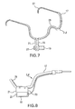
- FIG. 7 is a perspective view of a light therapy device according to an alternative embodiment in which an LED array is supported by a head-set.
- FIG. 7A is a perspective view of a light therapy device according to another alternative embodiment in which an LED array is supported by a head-set.
- FIG. 8 is a side view of the light therapy device of FIG. 7 .
- FIG. 9 is a front view of at least one LED array, and a connector detached from the head-set.
- FIG. 10 is a front view of an external light therapy device having two LED arrays, a hinge-like member, and an attaching means.
- FIG. 11 is a cross-sectional view of an LED array mounted onto a substrate.
- FIG. 11A is a schematic cross section through a portion of a light source having a light emitter and a reflector.
- FIG. 12 is a cross-sectional view of an LED array detached from the substrate.
- FIG. 1 shows an example light therapy apparatus 2 that comprises an extra-oral light source 4 having a right side 1 and a left side 3 (as viewed from the front of the device), an extra-oral bridge 5 , and an intra-oral tray 7 .
- Intra-oral tray 7 registers to a patient's teeth.
- Light source 4 is rigidly connected to intra-oral tray 7 by extra-oral bridge 5 . Therefore, a patient can position light source 4 accurately and repeatedly to illuminate a desired location in the patient's dental and maxillofacial areas by inserting intra-oral tray 7 into his or her mouth and biting intra-oral tray 7 so that it registers to at least some of the patient's teeth. This stabilizes light therapy apparatus 2 and positions light source 4 at a desired position.
- the consistent alignment and targeting of light from light source 4 during subsequent treatments makes the treatments more repeatable.
- extra-oral bridge 5 is removable from extra-oral light source 4 and intra-oral tray 7 .
- Providing a light therapy apparatus 2 having major components that are detachably connectable to one another adds versatility.
- a design which permits the major components of the light therapy apparatus to be disassembled and reassembled while preserving alignment of extra-oral light source 4 to intra-oral tray 7 has the advantage that the apparatus can be disassembled for storage or transportation and then used immediately after assembly.
- FIG. 3 shows light therapy apparatus 2 with extra-oral light source left side 3 detached from extra-oral bridge 5 .
- Extra-oral bridge 5 , extra-oral light source right side 1 , and extra-oral light source left side 3 may be secured together via a suitable connector.
- extra-oral bridge 5 , the extra-oral light source right side 1 , and the extra-oral light source left side 3 may be connected by inserting male connector portions 6 A of the extra-oral light source right and left sides 1 and 3 into corresponding female connector portions 8 A of extra-oral bridge 5 (see FIG. 3 ).
- the suitable connector allows extra-oral light source right and left sides 1 and 3 to be detached from extra-oral bridge 5 for ease of use and flexibility.
- extra-oral light source right and left sides 1 and 3 are rotatable between a sagittal orientation (as shown in FIG. 1 ) and a vertical orientation (indicated in dotted outline in FIG. 1 ).
- Light source right and left sides 1 and 3 can be locked at a desired angle of rotation by any suitable mechanism. This permits light source right and left sides 1 and 3 to be arranged so that the light that they emit fully covers the desired treatment areas.
- Intra-oral tray 7 may be connected to extra-oral bridge 5 by way of another suitable connector.
- a male portion 6 B of intra-oral tray 7 is removably received in a female portion 8 B of extra-oral bridge 5 .
- extra-oral bridge 5 may be reused for other patients (after suitable sterilization).
- Intra-oral tray 7 may be disposed of after it is no longer required by a patient.
- extra-oral bridge 5 is non-removably attached to intra-oral tray 7 .
- Intra-oral tray 7 is intended for insertion into a patient's mouth and is suitably shaped to fit around a patient's teeth. Intra-oral tray 7 may register with a few selected teeth (for example, intra-oral tray 7 may comprise a bite tab) or may fit around the patient's full set of teeth.
- the intra-oral tray 7 comprises a frame of a plastic or other suitable material that can serve as a skeleton for a settable material. The frame may be perforated to aid retention of the settable material.
- the frame may comprise extra-oral bridge 5 or a connector to connect to extra-oral bridge 5 .
- the frame for intra-oral tray 7 may be filled with a suitable settable material (for example a clear vinyl siloxane gel or similar material) which sets around the patient's teeth and subsequently allows repeatable alignment of intra-oral tray 7 in the patient's mouth.
- a suitable settable material for example a clear vinyl siloxane gel or similar material
- intra-oral tray 7 could be in the path of light as it travels from light source 4 to target tissues, the material of intra-oral tray 7 should be transparent to the light.
- Extra-oral bridge 5 preferably conforms around the jaw line of a patient.
- the light source right and left sides 1 and 3 are respectively positioned on the right and left sides of a patient's face along the patient's jaw line.
- Extra-oral bridge 5 may be adjustable to permit alignment of light source left and right sides 1 and 3 with target areas to be irradiated.
- Light source left and right sides 1 and 3 are extra-oral (outside of the patient's oral cavity). Light can pass from left and right sides 1 and 3 through tissues of the patient's lips and cheeks into target areas on the patient's gums and/or in the patient's jaws.
- Light source 4 emits light toward the patient.
- the light is not necessarily visible light.
- the light may include or consist of infrared light.
- Light source 4 comprises an array of light-emitting diodes (LEDs) in some embodiments.
- Light source 4 has an inner surface 13 (see FIG. 4 ) that is placed near or against the patient's skin adjacent to the tissues that it is desired to treat. Light is emitted is from inner surface 13 toward the area of treatment.
- left and right sides 1 and 3 of light source 4 each have a length similar to a significant fraction of the length of a human jaw.
- left and right sides 1 and 3 may each have a length of about 20 mm to about 90 mm in some embodiments and about 25 to about 45 or 60 mm in some embodiments.
- light source 4 may be smaller in extent.
- light source 4 has optics that emit light in the form of diverging beams.
- light source 4 may be somewhat smaller than the area of tissues to be treated because light from light source 4 will spread somewhat as it passes through the tissues of the patient's lips and cheeks before reaching the tissues of the jaw and or gums to be treated.
- Light source 4 may be wide enough to illuminate both upper and lower jaws of a patient simultaneously although in some embodiments light source 4 may be narrower.
- light source 4 has a width in the range of 12 mm to about 40 mm in some embodiments (e.g. about 15 to 17 mm in some embodiments).
- the light is emitted by arrays of discrete LEDs.
- the LEDs may be arranged in any of a wide variety of patterns.
- the LEDs may be arranged in staggered parallel rows to maximize the density of LEDs in the LED array.
- the LEDs may be arranged to achieve substantially uniform optical intensity over the light-emitting inner surface 13 of light source 4 .
- each array comprises 50 to 100 LEDs or other light emitters.
- the average light intensity produced by light source 4 is at least about 10 mW/cm 2 .
- light source 4 has an average intensity that is, or can be adjusted to be, in the range of 20 mW/cm 2 to about 60 mW/cm 2 .
- the output of light source 4 is pulsed.
- the peak light intensity may be significantly higher than 50 mW/cm 2 .
- right light source 4 or its components are flexible so that they can be bent in one or two dimensions (i.e. molded) to conform to the contours of the patient's face.
- light source 4 may comprise an array of light emitters mounted to a flexible sheet of material that will hold a shape when it is bent.
- the flexible material can advantageously comprise a metal sheet that can serve as a heat sink or as a thermal path to a heat sink for heat generated by the light emitters.
- the flexible sheet may be molded to conform to the contours of the patient's face while light therapy apparatus 2 is being fitted.
- Light source 4 may include optical elements such as lenses and reflectors to focus and direct light from light source 4 onto a target area. Such optical elements may be suitably encapsulated in plastic or similar material.
- FIG. 11A shows a portion of a light source 4 .
- a light emitter 11 (which may, for example, comprise a junction in a light-emitting diode or other light-emitting semiconductor device) is located adjacent to a reflective backing 11 A.
- a curved light-reflecting recess 11 B is provided adjacent to light emitter 11 . Light from light source 11 is reflected in recess 11 B to form a beam.
- the beams from all light emitters of light source 4 combine to illuminate the target tissues. The area covered by the beam will depend upon the tissues which it is desired to treat.
- the beams of light emitted by light source 4 diverges to cover an area of tissue larger than the area of the light-emitting part of light source 4 .
- the emitted light converges to provided increased light intensity at the location of the tissues that it is desired to treat.
- the emitted light diverges in a beam having an included angle ⁇ in the range of about 45-60°.
- light source 4 may comprise a system for forced air or liquid cooling.
- a cooling system allows for treatment without the danger of potential burns to the patient and allows for greater efficiency and control of the device.
- Extra-oral light source right and left sides 1 and 3 may comprise thermally-conductive LED wafers mounted on a suitable heat sink. Heat from the LED wafers is conducted into the heat sink and dissipated.
- FIGS. 1A , 1 B and 1 C show a light source 100 of a type that may be used as light source right and left sides 1 and 3 .
- Light source 100 comprises arrays 102 of LEDs that are mounted to a heat sink 104 .
- Heat sink 104 has pins 106 projecting from its face that is away from LED arrays 102 .
- a fan 110 causes air to flow past pins 106 to carry away excess heat.
- the light from light source 4 at the tissues to be treated should have at least a threshold intensity.
- Light source 4 may be operated in a pulsed mode to facilitate cooling of light source 4 while ensuring that when light source 4 is emitting light, the intensity of emitted light at the tissues to be treated is sufficient to be effective.
- the duty cycle of light source 4 is 1:1 or less, in some embodiments 1:2 or less (for each interval in which light source 4 is on, light source 4 is off for two equal intervals).
- the pulsing of light source 4 may be performed fast enough that light source 4 does not visibly flicker (e.g. at 25 Hz or more) although this is not mandatory.
- the character of the light emitted by light source right and left sides 1 and 3 will depend upon the nature of the LEDs or other light emitters in light source 4 . It is generally desirable that the emitted light include light in the wavelength range of 620 nm to 1000 nm. In some embodiments the emitted light includes light having a wavelength in at least one of the following wavelength ranges: about 820 to about 890 nm and about 620 to about 680 nm. Light having wavelengths corresponding to one or more of the following ranges may be particularly effective:
- the light is substantially monochrome in some embodiments although this is not mandatory. Providing light emitters that emit at multiple wavelengths allows for irradiation over multiple wavelengths for greater biological activity.
- the light may comprise incoherent light although this is not mandatory.
- the light may be delivered continuously or pulsed at suitable frequencies and duty cycles.
- Invisible infrared light can be clinically effective.
- the emitted light includes infrared light
- the emitted light also includes bright visible light.
- the bright visible light deters users from looking into the light source when it is operating, provides a perceptible indication that the apparatus is operating, and may be useful in properly positioning the device.
- the visible light may be, but is not necessarily in a wavelength range that is beneficial for light therapy.
- the ratio of the intensities of the visible and infrared components of the light is 1 part or less visible light to 5 parts or more infrared light.
- the treatment area and desired light characteristics will vary from patient to patient.
- a physician, dentist or other therapist can determine a light treatment regime for a patient and set up light therapy apparatus 2 to operate light emitters in light source 4 to provide the desired treatment.
- FIG. 6 illustrates a programmable controller 15 of a type that may be used to control the operation of light therapy apparatus 2 (or other light therapy apparatus as described below).
- Programmable controller 15 may be a separate, remote unit or may be directly connected to or integrated with light source 4 .
- Programmable controller 15 may comprise a microprocessor, data store, power supply, clock and associated electronic circuitry. Control parameters are stored in the data store. Programmable controller 15 operates light source 4 according to the parameters in the data store. The parameters may specify one or more of:
- light therapy apparatus has sets of light emitters having different characteristics (e.g. sets of LED that emit light at different wavelengths or sets of light emitters that illuminate target tissues in different locations) then separate control parameters may be provided for different sets of the light emitters.
- different sets of parameters are specified for different segments (intervals) of a light treatment.
- light therapy treatments may be defined for a set of intervals each lasting from a few seconds to a few hundred seconds or a fraction of an hour. Different parameters may be specified for each of the intervals. The intervals are not necessarily equal in length.
- different sets of parameters may be specified for different areas of light source 4 .
- some areas of light source 4 may be turned off because the treatment plan for a patient does not require light to be delivered at locations corresponding to those parts of the light source 4 .
- a physician, dentist, or therapist may program a patient's treatment regimen into programmable controller 15 . This may be done, for example, with the aid of suitable software running on a computer that is in data communication with programmable controller 15 or by way of a suitable user interface built into programmable controller 15 .
- Programmable controller 15 may have one or more pre-set programs built in. As an alternative to, or as an aid to programming controller 15 the physician, dentist, or therapist may select a pre-set program that is appropriate for controlling light therapy apparatus 2 to deliver light to a patient.
- a typical treatment regimen provides a dose of light daily.
- Each of the daily doses of light may be delivered over a period lasting between a few minutes and an hour or so.
- daily 1 ⁇ 2 hour doses of light can be effective and are not unduly inconvenient for patients.
- a single daily dose appears to be as effective as dividing the same dose into multiple sessions delivered at different times during the day. Examples of possible treatment regimens are:
- Programmable controller 15 may maintain a log of treatments that have been delivered. For example, controller 15 may log the date and time that each treatment was initiated, the duration of the treatment, and whether or not the treatment was completed. This log can be subsequently reviewed by a dentist, physician, or the like to evaluate whether or not the patient has complied with the prescribed treatment regimen.
- Programmable controller 15 has a button or other suitable user patient interface that allows a patient to initiate a treatment according to previously-set parameters in the data store.
- the patient interface is preferably very simple such that minimal instruction is required to explain to a patient how to use light therapy apparatus 2 .
- Programmable controller 15 may include an audible or visual indicator that generates a signal to remind a patient that it is time for a treatment (or that a scheduled treatment is overdue).
- a patient can use light therapy apparatus 2 at home or in another location by operating programmable controller 15 to initiate delivery of a treatment.
- Programmable controller 15 may comprise circuitry that monitors temperature at one or more locations in light source 4 .
- the circuitry may monitor a signal modulated by a temperature sensor in light source 4 .
- programmable controller 15 may monitor the current and voltage driving LEDs in light source 4 .
- the current/voltage relationship is temperature-dependent. Thus, by monitoring the current/voltage relationship programmable controller 15 can determine whether the LED is at an undesirably high temperature.
- Programmable controller 15 may shut off or reduce current to light source 4 (or part of light source 4 ) when it detects that the temperature of light source 4 is undesirably high (or is trending towards being undesirably high). If light source 4 is equipped with a cooling fan then programmable controller 15 may optionally control the speed of the cooling fan in response to the monitored temperature.
- Programmable controller 15 may be configured to maintain a log of treatments delivered by light therapy apparatus 2 .
- the log may be reviewed by a physician, dentist or technician to verify that light therapy device has been used as prescribed by a patient.
- the log may track the times and durations of light therapy treatments delivered by light therapy apparatus 2 and may also track other features such as operating temperatures, operational status and the like.
- FIGS. 7 and 8 show a light therapy apparatus 2 A having a head-set style arrangement.
- Light therapy apparatus 2 A comprises a head-set 17 and at least one extra-oral light source 19 mounted to head-set 17 by way of a suitable connector 21 .
- Head-set 17 may have the general form of a frame for eyeglasses.
- headset 17 has arms 27 that fit above and around the patient's ears and a frame 29 that fits over the bridge of the patient's nose.
- Head-set 17 may also include lenses (not shown).
- the lenses may be made of a material that blocks radiation at wavelengths emitted by light source 19 so that the patient's eyes are protected from the radiation.
- Light source 19 may comprise an array of LEDs or other light emitters.
- Head-set 17 When head-set 17 has been adjusted to fit an individual patient, frame 29 registers with the bridge of the patient's nose and arms 27 sit on the patient's ears. Head-set 17 will sit on the patient's head in the same way each time it is put on. Head set 17 may be adjusted for fit by adjusting arms 27 which may be made of a firm, resilient material that allows for some flexibility for a better and more secure fit for individual users. In some embodiments, arms 27 can also be adjusted horizontally along their axis. Frame 29 can also be adjustable, for example, by bending to allow for a better and more secure fit. An elastic keeper such as an elastic strap may be provided to hold head-set 17 in place during use.
- arms 27 may be made of a firm, resilient material that allows for some flexibility for a better and more secure fit for individual users.
- arms 27 can also be adjusted horizontally along their axis.
- Frame 29 can also be adjustable, for example, by bending to allow for a better and more secure fit.
- An elastic keeper such as an
- connector 21 permits the position of light source 19 to be adjusted both along a horizontal axis 30 A and a vertical axis 30 B relative to head-set 17 .
- a yoke 31 A is mounted to head-set 17 by screws 31 B which pass through slot 31 C.
- the position of light source 19 in horizontal direction 30 A can be adjusted by loosening screws 31 B, sliding yoke 31 A to a desired position along slot 31 C and retightening screws 31 B.
- Light source 19 is connected to arms 31 D of yoke 31 A by screws 31 E which pass through slots 31 F.
- the vertical position of light source 19 may be adjusted by loosening screws 31 E, sliding light source 19 up or down along slots 31 F to a desired vertical position and then retightening screws 31 E.
- slot 31 C is curved when viewed from above.
- Slot 31 C generally follows the curvature of a typical maxillary bone such that light source 19 can effectively be applied against the patient's skin for a range of positions of light source 19 along slot 31 C.
- connector 21 may hold light source 19 so that it is tilted with its lower edge projecting more in the direction of the patient than its upper edge.
- the angle of tile of light source 19 is adjustable. Head-set 17 may be adjusted so that light source 19 is biased against the patient's face when head set 17 is being worn by a patient.
- connector 21 may comprise a bar, rod or similar device that can be clamped or otherwise fastened to head-set 17 and a clip or similar mechanism that fastens light source 19 to the bar, rod or similar device.
- light source 19 can be removably detached from head-set 17 . This can be convenient for storage or transportation of light therapy apparatus 2 A.
- head-set 17 comprises an adjustable strap (not shown) which fits around the crown of a patient's head for securing the extra-oral light therapy device 2 A.
- the adjustable strap can also fit around a patient's chin and extend back to the crown and around the crown of a patient's head.
- the adjustable strap may be made of a flexible, elastic woven material.
- FIG. 10 shows a light therapy apparatus 34 comprising at least one light source 35 .
- Light source 35 comprises at least one light emitter, for example an LED array, mounted on a thin molded substrate 51 ( FIG. 11 ). More than one array of light emitters may be provided in light source 35 .
- the light source 35 shown in FIG. 10 has two arrays of LEDs.
- Arrays 36 of light emitters may be arranged in lower level 45 and an upper level 47 .
- the upper and lower levels may be separately controlled.
- the upper and lower levels respectively irradiate tissues of the upper and lower jaws.
- An attaching means 43 is provided for securing the device to the area of treatment.
- a power source and controller which may comprise a programmable controller 15 as described above, operate light source 35 to emit light according to a desired protocol.
- light source 35 has a right section 37 , a center section 39 and a left section 41 .
- Right section 37 and the left section 41 are respectively supported on the right and left sides of a patient's face.
- a light source 35 as shown in FIG. 10 may be supported by way of any suitable attaching means including:
- the LED arrays may be removably attached to light source 35 by suitable connectors 38 such as ribbon connectors or may be more permanently integrated into light source 35 as illustrated in FIG. 11 .
- suitable connectors 38 such as ribbon connectors or may be more permanently integrated into light source 35 as illustrated in FIG. 11 .
- Providing removable, repositionable LED arrays on a light source 35 permits LED arrays to be arranged on light source 35 so as to optimally illuminate target tissues. LED arrays may be concentrated to illuminate target tissues while areas of light source 35 that overly non-target tissues do not need to have any LED arrays.
- FIG. 12 shows a cross-section of an LED array 36 of external light therapy device 34 detached from substrate 51 .
- a clip or similar attaching means 53 allows the at least one LED array 36 to be mounted onto substrate 51 .
- Substrate 51 serves as a heat sink as described above.
- Substrate 51 may be made of aluminum or similar metal that is a good heat conductor.
- Substrate 51 may be moldable (i.e. flexible in one or two dimensions so that it can be formed to follow contours of a patient's face and, once formed, will retain its shape).
- Hinge-like members 49 may be provided between arrays 36 to allow light source 35 to be bent to provide a better fit around the facial area.
- Hinge-like member 49 may comprise a thin crease 50 or other bend line set into the substrate material, as illustrated in FIG. 11 .
- Hinge-like member 49 allows the center section 39 to fit around a patient's mouth and the right section 37 and the left section 41 to fit around a patient's face.
- Apparatus as described herein may be applied to treat a variety of conditions including:
- the apparatus may be applied by fitting a support to a patient.
- the support may comprise a head-set, intra-oral tray, a bite tab, or the like.
- one or more light sources are mounted to the support at locations where light from the light sources can illuminate a treatment area.
- a treatment regimen is then established.
- the physician, dentist, or therapist at his office or a patient at his home then performs the prescribed extra-oral light therapy treatment.
- intra-oral tray 7 Prior to extra-oral light therapy treatment, intra-oral tray 7 is prepared by filling it with a suitable settable material such as a clear vinyl siloxane gel or similar material. The intra-oral tray is then placed around the patient's teeth to obtain an impression of the patient's teeth. After the settable material sets, intra-oral tray 7 can be used to achieve consistent targeting of light to target tissues bone during subsequent treatments.
- a suitable settable material such as a clear vinyl siloxane gel or similar material.
- a physician, dentist, or therapist programs a patient's prescribed treatment regimen into a programmable controller 15 (see FIG. 6 , for example).
- Programmable controller 15 controls parameters of a light therapy treatment to be delivered by light therapy apparatus 2 .
- controller 15 may control the duration of the treatment, light intensity, pulse frequency, etc.
- Programmable controller 15 runs a patient's prescribed treatment regimen causing the at least one light source 4 to emit pulsed or continuous light according to the prescribed parameters onto the treatment area. Therefore, stimulating and accelerating bone formation and healing at a patient's treatment area for the treatment of jaw bone disorders and jaw osteonecrosis.
- the invention also relates to a method for the treatment and stimulation of soft and hard tissue and the biostimulation of bone.
- a light source 35 which may comprise at least one LED array 36 is first attached to the desired area of treatment.
- a physician, dentist, or therapist programs a patient's prescribed treatment regimen into a programmable controller 15 .
- Programmable controller 15 controls the energy density, pulse frequency and duration of the external light therapy device 34 .
- the programmable controller 15 runs a patient's prescribed treatment regimen causing the at least one LED array 35 to emit pulsed or continuous light at the predetermined rates and frequencies onto the treatment area.
- the light therapy device can provide effective, stabilized, repeatable, accurate, programmable, and consistent light therapy for the treatment and stimulation of soft and hard tissue and the biostimulation of bone.
- Exclusion criteria included: 1) a medical condition associated with abnormal bone growth or remodeling, such as Paget's disease of bone, fibrous dysplasia, osteopetrosis, severe systemic osteoporosis, etc.; 2) unwillingness to sign informed consent form; 3) inability to perform daily LED treatments at home; 4) inability to obtain high quality QUS scans of the jaws.
- An exclusion waiver for the research was provided by the Committee for the Protection of Human Subjects of the University of Texas in Houston and informed consent was obtained from all subjects.
- III Cube shows moderate loss of column height in more than 1 ⁇ 2 of columns (32 columns); and/or severe loss of column height in 1 ⁇ 4 to 1 ⁇ 2 of columns (17-32 columns).
- IV Cube shows severe loss of column height in more than 1 ⁇ 2 of columns (32 columns).
- the investigational OsseoPulseTM (Version 1.0) device made by Biolux Research Ltd., Vancouver, Canada.
- the device consists of an extra-oral array of highly-efficient light emitting diodes (LED) producing non-coherent continuous wave monochromatic light in the visible far red (660 nm @ 15 mW/cm2) and infra-red range (840 nm @ 20 mW/cm2).
- LED highly-efficient light emitting diodes
- the OsseoPulse device was placed on the facial surface for 15 minutes daily, 5 days a week for 12 weeks on each treatment side.
- the dose per session per treatment area was approximately 200 Joules per square inch.
Abstract
Description
-
- 613 nm to 624 nm
- 667 nm to 684 nm
- 750 nm to 773 nm
- 812 nm to 846 nm.
The range 613 nm to 624 nm corresponds to a band at which reduced cytochrome c oxidase absorbs light. The range 812 nm to 846 nm corresponds to a band at which oxidized cytochrome c oxidase absorbs light.
-
- treatment duration;
- light intensity during the treatment;
- whether light emitters operate continuously or are pulsed;
- if the light emitters are pulsed, the rate at which light emitters are pulsed;
- if the light emitters are pulsed, the duty cycle at which the light emitters are pulsed;
- etc.
-
- Enhancement of bone density by applying light in 5 treatments per week for 12 weeks. Each treatment lasts ½ hour and illuminates the tissues of a patient's jaw with light having wavelengths of 660 nm and 840 nm. The 660 nm light has an intensity of about 20 mW/cm2 at the skin's surface The 840 nm light has an intensity of about 10 mW/cm2 at the skin's surface.
- Accelerating healing of bone grafts by applying light in daily treatments for 21 days. Each treatment lasts between 20 minutes and one hour and illuminates the tissues of a patient's jaw with light having a wavelength of 618 nm and an intensity of 20 mW/cm2 at the skin's surface.
-
- a head-set 17 as described above;
- an
intra-oral tray 7 which may comprise a full tray or one or more bite tabs as described above; - an adhesive such as double-sided adhesive tape;
- a strap or set of straps; or
- the like.
-
- jaw osteonecrosis,
- other jaw bone disorders,
- periodontitis,
- malocclusion and other conditions treated by orthodontics,
- stimulation and acceleration of healing after oral surgery or periodontal surgery,
- stimulation of the healing of wounds at the locations of bone grafts,
- healing and acceleration of osseo-integration of endosseous dental implants; and,
- the like.
The application to jaw osteonecrosis permits treatment of a condition for which existing treatments are highly invasive. Treating osteonecrosis using light therapy is significantly more cost-effective and comfortable for the patient than existing surgical treatment options.
| TABLE 1 |
| Grading categories for individual 3-D cube images (64 columns in each) |
| of the Cavitat QUS images. |
| QUS | |
| Grade * | Description ** |
| 0 | “Green bone.” Cube shows no loss of column height and is |
| 100% green; or mild loss of column height in less than ¼ of | |
| columns (16 columns); and/or moderate to severe loss of | |
| column height in less than 4 non-adjacent columns. | |
| I | Cube shows mild loss of column height in more than ¼ of |
| columns; and/or moderate loss of column height in 1/16 to | |
| ¼ of the columns (5-16 columns); and/or severe loss of | |
| height in 1/16 to ⅛ of the columns (5-8 columns). | |
| II | Cube shows moderate loss of column height in ¼ to ½ of |
| columns (17-32 columns); and/or severe loss of height in ⅛ to | |
| ¼ of columns (8-16 columns). | |
| III | Cube shows moderate loss of column height in more than ½ of |
| columns (32 columns); and/or severe loss of column height in | |
| ¼ to ½ of columns (17-32 columns). | |
| IV | Cube shows severe loss of column height in more than ½ of |
| columns (32 columns). | |
| * high grade lesion = Grade III and IV scans; low-grade lesion = Grade I and II scans; “green bone” = normal or Grade 0 scan | |
| ** definition of loss of column height: mild (crown is green, less than ⅓ loss of height); moderate (crown is yellow or brown, ⅓ to ⅔ loss of height); severe (crown is orange or red, more than ⅔ loss of height) | |
| TABLE 2 |
| Results of 294 QUS scans before and after 3 months |
| of daily LED photobiomodulation. |
| Grade Level * | # at Pre-Treatment | # at Post-Treatment | |
| 1 | 79 | 120 | |
| 2 | 69 | 54 | |
| 3 | 86 | 53 | |
| 4 | 61 | 40 | |
| Mean: | 2.43 | 1.33 | |
| * 1 = mild LBD/dehydration; 4 = severe LBD/dehydration | |||
| TABLE 3 |
| Post-treatment changes for each pre-treatment grade level, 294 QUS scans. |
| Number of Sites | Number @ Grade for each Site at | ||
| Grade | at | Post-Treatment* | Avg. |
| Level* | Pre-Treatment | 0** | 1** | 2** | 3** | 4** | |
| 1 | 79 | 54 | 15 | 8 | 2 | 0 | −0.54 |
| 2 | 69 | 32 | 19 | 12 | 4 | 2 | −1.32 |
| 3 | 86 | 26 | 17 | 22 | 16 | 5 | −1.50 |
| 4 | 60 | 8 | 3 | 10 | 18 | 21 | −1.32 |
| Mean: | 2.43 | 120 | 54 | 52 | 40 | 28 | −1.11 |
| *1 = mild LBD/dehydration; 4 = severe LBD/dehydration (see Table 1) | |||||||
| **Represents grade levels, 0-4, as described in Table 1 | |||||||
-
-
Light therapy apparatus 34 may be applied for treatment and stimulation of other bone or soft tissues, such as the hip. In such applications,light source 35 can be attached to a treatment area with an adhesive such as double-sided adhesive tape (not shown). Alternatively, the externallight therapy apparatus 34 can be placed or sewn into a pouch, undergarment or similar garment and attached to the treatment area through means of a strap, button or similar attaching means (not shown). - It is not mandatory that a controller be programmable. For example, a controller may have controls that allow various parameters to be set. A physician, therapist or technician may set those controls so that an appropriate treatment is delivered when a patient initiates delivery of the treatment.
- Features or components described in relation to one of the embodiments described herein may be provided in combination with components or features of other ones of the example embodiments described herein. For example, the
controller 15 shown inFIG. 6 could be used in conjunction with any of the described embodiments. Light sources having a property or properties like those of thelight source 4 shown in the embodiments ofFIGS. 1 to 1C could be applied in other embodiments.
It is therefore intended that the following appended claims and claims hereafter introduced are interpreted to include all such modifications, permutations, additions and sub-combinations as are within their true spirit and scope.
-
Claims (36)
Priority Applications (2)
| Application Number | Priority Date | Filing Date | Title |
|---|---|---|---|
| US13/895,330 US8900282B2 (en) | 2005-02-17 | 2013-05-15 | Light therapy apparatus and methods |
| US14/554,404 US9308389B2 (en) | 2005-02-17 | 2014-11-26 | Light therapy apparatus and methods |
Applications Claiming Priority (5)
| Application Number | Priority Date | Filing Date | Title |
|---|---|---|---|
| US65382805P | 2005-02-17 | 2005-02-17 | |
| US70575305P | 2005-08-05 | 2005-08-05 | |
| US11/355,583 US20060200212A1 (en) | 2005-02-17 | 2006-02-16 | Light therapy device for treatment of bone disorders and biostimulation of bone and soft tissue |
| US11/767,302 US20070248930A1 (en) | 2005-02-17 | 2007-06-22 | Light therapy apparatus and methods |
| US13/895,330 US8900282B2 (en) | 2005-02-17 | 2013-05-15 | Light therapy apparatus and methods |
Related Parent Applications (1)
| Application Number | Title | Priority Date | Filing Date |
|---|---|---|---|
| US11/767,302 Continuation US20070248930A1 (en) | 2005-02-17 | 2007-06-22 | Light therapy apparatus and methods |
Related Child Applications (1)
| Application Number | Title | Priority Date | Filing Date |
|---|---|---|---|
| US14/554,404 Division US9308389B2 (en) | 2005-02-17 | 2014-11-26 | Light therapy apparatus and methods |
Publications (2)
| Publication Number | Publication Date |
|---|---|
| US20130253620A1 US20130253620A1 (en) | 2013-09-26 |
| US8900282B2 true US8900282B2 (en) | 2014-12-02 |
Family
ID=40193520
Family Applications (5)
| Application Number | Title | Priority Date | Filing Date |
|---|---|---|---|
| US11/767,302 Abandoned US20070248930A1 (en) | 2005-02-17 | 2007-06-22 | Light therapy apparatus and methods |
| US12/834,601 Abandoned US20100318161A1 (en) | 2005-02-17 | 2010-07-12 | Light therapy methods |
| US13/895,330 Expired - Fee Related US8900282B2 (en) | 2005-02-17 | 2013-05-15 | Light therapy apparatus and methods |
| US14/147,210 Abandoned US20140121731A1 (en) | 2005-02-17 | 2014-01-03 | Light therapy methods |
| US14/554,404 Active US9308389B2 (en) | 2005-02-17 | 2014-11-26 | Light therapy apparatus and methods |
Family Applications Before (2)
| Application Number | Title | Priority Date | Filing Date |
|---|---|---|---|
| US11/767,302 Abandoned US20070248930A1 (en) | 2005-02-17 | 2007-06-22 | Light therapy apparatus and methods |
| US12/834,601 Abandoned US20100318161A1 (en) | 2005-02-17 | 2010-07-12 | Light therapy methods |
Family Applications After (2)
| Application Number | Title | Priority Date | Filing Date |
|---|---|---|---|
| US14/147,210 Abandoned US20140121731A1 (en) | 2005-02-17 | 2014-01-03 | Light therapy methods |
| US14/554,404 Active US9308389B2 (en) | 2005-02-17 | 2014-11-26 | Light therapy apparatus and methods |
Country Status (3)
| Country | Link |
|---|---|
| US (5) | US20070248930A1 (en) |
| EP (1) | EP2164570A4 (en) |
| WO (1) | WO2009000075A1 (en) |
Cited By (10)
| Publication number | Priority date | Publication date | Assignee | Title |
|---|---|---|---|---|
| US20150079536A1 (en) * | 2005-02-17 | 2015-03-19 | Biolux Research Ltd. | Light therapy apparatus and methods |
| US20170224455A1 (en) * | 2013-08-01 | 2017-08-10 | Jbl Radical Innovations, Llc | Closed system mouthpiece with light and heat generation to activate a formulation to increase its volume |
| US9730780B2 (en) | 2013-10-22 | 2017-08-15 | Biolux Research Ltd. | Intra-oral light-therapy apparatuses and methods for their use |
| US10569097B2 (en) | 2015-07-28 | 2020-02-25 | Photonmd, Inc. | Systems and methods for phototherapeutic modulation of nitric oxide |
| US10688315B2 (en) | 2015-07-28 | 2020-06-23 | Know Bio, Llc | Phototherapy devices for treatment of dermatological disorders of the scalp |
| US11147984B2 (en) | 2020-03-19 | 2021-10-19 | Know Bio, Llc | Illumination devices for inducing biological effects |
| US11285337B2 (en) | 2016-07-22 | 2022-03-29 | The Regents Of The University Of Michigan | Temporally modulated multi-LED for enhanced subconscious physiological responses |
| US11395719B2 (en) | 2018-11-26 | 2022-07-26 | Anthony T Suriano | Unimpeded distalizing jig |
| US11458329B2 (en) | 2016-07-27 | 2022-10-04 | Z2020, Llc | Componentry and devices for light therapy delivery and methods related thereto |
| US11654294B2 (en) | 2021-03-15 | 2023-05-23 | Know Bio, Llc | Intranasal illumination devices |
Families Citing this family (47)
| Publication number | Priority date | Publication date | Assignee | Title |
|---|---|---|---|---|
| US20060200212A1 (en) * | 2005-02-17 | 2006-09-07 | Brawn Peter R | Light therapy device for treatment of bone disorders and biostimulation of bone and soft tissue |
| EP2339983B1 (en) * | 2008-09-09 | 2014-11-26 | New York University | Devices to increase craniofacial bone density |
| WO2010142013A1 (en) * | 2009-06-08 | 2010-12-16 | Biolux Research Limited | Method and device for accelerating orthodontic tooth movement |
| US9642687B2 (en) | 2010-06-15 | 2017-05-09 | The Procter & Gamble Company | Methods for whitening teeth |
| EP2627283B1 (en) * | 2010-10-13 | 2015-09-23 | Biolux Research Limited | Apparatus for tooth regulation with heavy forces |
| EP2640461B1 (en) | 2010-11-16 | 2019-06-19 | The Board Of Trustees Of The Leland Stanford Junior University | Systems for treatment of dry eye |
| US9821159B2 (en) | 2010-11-16 | 2017-11-21 | The Board Of Trustees Of The Leland Stanford Junior University | Stimulation devices and methods |
| US9242118B2 (en) * | 2010-12-08 | 2016-01-26 | Biolux Research Ltd. | Methods useful for remodeling maxillofacial bone using light therapy and a functional appliance |
| CA2854923C (en) * | 2011-11-08 | 2021-10-19 | Biophotas, Inc. | Shapeable light therapy device and method |
| US9687669B2 (en) | 2011-11-09 | 2017-06-27 | John Stephan | Wearable light therapy apparatus |
| US9352170B1 (en) | 2012-01-31 | 2016-05-31 | Christina Davis | Spectral light therapy for autism spectral disorders |
| AU2013246421B2 (en) | 2012-04-13 | 2017-09-28 | Advanced Orthodontics And Education Association, Llc | Method and device for increasing bone density in the mouth |
| EP3626206A1 (en) * | 2012-04-19 | 2020-03-25 | Biolux Research Holdings, Inc. | Intra-oral light therapy apparatus |
| US20130280671A1 (en) * | 2012-04-19 | 2013-10-24 | Biolux Research Ltd. | Intra-oral light therapy apparatuses and methods for their use |
| US9717627B2 (en) | 2013-03-12 | 2017-08-01 | Oculeve, Inc. | Implant delivery devices, systems, and methods |
| WO2014172693A2 (en) | 2013-04-19 | 2014-10-23 | Oculeve, Inc. | Nasal stimulation devices and methods |
| CA2917724A1 (en) * | 2013-07-17 | 2015-01-22 | Meditech International Inc. | System and method for multi-colour light treatment |
| US9846311B2 (en) * | 2013-07-30 | 2017-12-19 | Jonathan Stephen Farringdon | Method and apparatus for forming a visible image in space |
| EP3110405B1 (en) | 2014-02-25 | 2020-05-06 | Oculeve, Inc. | Polymer formulations for nasolacrimal stimulation |
| AU358535S (en) | 2014-04-18 | 2014-11-03 | Oculeve | Nasal stimulator device |
| EP3673952A1 (en) | 2014-07-25 | 2020-07-01 | Oculeve, Inc. | Stimulation patterns for treating dry eye |
| KR101525123B1 (en) * | 2014-08-29 | 2015-06-03 | 주식회사 비에스앤코 | Teeth Whitening Apparatus |
| EP3209371A4 (en) | 2014-10-22 | 2018-10-24 | Oculeve, Inc. | Implantable nasal stimulator systems and methods |
| AU2015335776B2 (en) | 2014-10-22 | 2020-09-03 | Oculeve, Inc. | Stimulation devices and methods for treating dry eye |
| WO2016065211A1 (en) | 2014-10-22 | 2016-04-28 | Oculeve, Inc. | Contact lens for increasing tear production |
| US9844426B2 (en) * | 2015-03-12 | 2017-12-19 | Align Technology, Inc. | Digital dental tray |
| BE1022600B1 (en) * | 2015-04-29 | 2016-06-14 | Hugues Libotte | AUTONOMOUS DEVICE FOR VISIBLE AND INFRARED ILLUMINATION TREATMENT OF ARTHROSITIC PAIN IN SMALL ANIMALS |
| US10426958B2 (en) | 2015-12-04 | 2019-10-01 | Oculeve, Inc. | Intranasal stimulation for enhanced release of ocular mucins and other tear proteins |
| US10252048B2 (en) | 2016-02-19 | 2019-04-09 | Oculeve, Inc. | Nasal stimulation for rhinitis, nasal congestion, and ocular allergies |
| WO2017147599A1 (en) | 2016-02-26 | 2017-08-31 | Cimphoni Life Sciences LLC | Light emitting bone implants |
| CA3022683A1 (en) | 2016-05-02 | 2017-11-09 | Oculeve, Inc. | Intranasal stimulation for treatment of meibomian gland disease and blepharitis |
| USD860453S1 (en) * | 2016-10-10 | 2019-09-17 | K-Laser D.O.O. | Device for laser therapy |
| RU2019118600A (en) | 2016-12-02 | 2021-01-11 | Окулив, Инк. | APPARATUS AND METHOD FOR MAKING DRY EYE SYNDROME PREDICTION AND TREATMENT RECOMMENDATIONS |
| JP3209729U (en) * | 2017-01-23 | 2017-04-06 | 秀俊 西尾 | Light irradiation device for whitening |
| CN110325142B (en) | 2017-01-25 | 2022-02-01 | 登士柏希罗纳有限公司 | Light-cured dental system |
| US11202919B2 (en) | 2017-03-30 | 2021-12-21 | Healthe, Inc. | Wavelength converting therapeutic treatment and associated methods |
| US11141254B2 (en) | 2018-08-10 | 2021-10-12 | Sdc U.S. Smilepay Spv | Mouthpiece for teeth whitening |
| US10716652B2 (en) | 2018-08-10 | 2020-07-21 | SDU U.S. SmilePay SPV | Mouthpiece for teeth whitening |
| US11040218B2 (en) * | 2018-10-11 | 2021-06-22 | Colgate-Palmolive Company | Oral treatment device, system and method |
| US20230210453A1 (en) | 2018-11-28 | 2023-07-06 | Biolux Research Holdings Inc | Orthodontic appliance compliance monitoring systems, devices, and methods |
| USD925046S1 (en) | 2019-05-30 | 2021-07-13 | Biophotas, Inc. | Light therapy device |
| US11331513B2 (en) | 2019-09-07 | 2022-05-17 | National Laser Company | Pain treatment device |
| US20210128280A1 (en) * | 2019-11-01 | 2021-05-06 | Millennium Healthcare Technologies, Inc. | Blast protocol |
| USD1008700S1 (en) | 2021-02-08 | 2023-12-26 | Jonathan A. Shanker | Neck therapy cushion |
| US11833368B2 (en) | 2020-02-11 | 2023-12-05 | Jonathan A. Shanker | Systems and methods for delivering low-level electromagnetic radiation to a patient |
| USD957660S1 (en) | 2020-02-28 | 2022-07-12 | Biophotas, Inc. | Controller for light therapy system |
| CN113995960B (en) * | 2021-11-23 | 2023-12-08 | 固安翌光科技有限公司 | Oral cavity internal light medical device |
Citations (176)
| Publication number | Priority date | Publication date | Assignee | Title |
|---|---|---|---|---|
| US2635175A (en) | 1952-02-09 | 1953-04-14 | Hodge Woodrow Wilson | Therapeutic appliance |
| US2884926A (en) | 1957-03-15 | 1959-05-05 | Grasso Frank | Device for radiating the mouth |
| US3516411A (en) | 1968-05-13 | 1970-06-23 | Estelle Adler | Apparatus for the therapeutic treatment of the skin |
| US3971387A (en) | 1975-03-21 | 1976-07-27 | Mantell Michael J | Electro-therapeutic face mask |
| US4244373A (en) | 1978-05-17 | 1981-01-13 | Nachman Marvin J | Electrical stimulation dental device |
| US4273535A (en) | 1978-12-04 | 1981-06-16 | Kabushiki Kaisha Morita Seisakusho | Device for preventing tooth decay by laser beam irradiation and method of preventing tooth decay by use of the same |
| US4457707A (en) | 1981-11-18 | 1984-07-03 | Medical Magnetics, Inc. | Integrated oral magnetic osteogenic and orthodontic appliances |
| US4628931A (en) | 1982-03-03 | 1986-12-16 | Barrett Harold F | Medical treatment method |
| GB2203649A (en) | 1987-04-15 | 1988-10-26 | Kei Mori | Light radiation device |
| US4840174A (en) | 1986-06-27 | 1989-06-20 | University Of Cincinnati | Method of laser treatment of cancerization of the oral cavity |
| GB2212010A (en) | 1987-11-04 | 1989-07-12 | Amcor Ltd | Radiation therapy apparatus using LED matrix |
| US4877401A (en) | 1988-03-09 | 1989-10-31 | University Of Utah | Method of preventing tooth decay by laser beam irradiation and chemical treatment |
| US4983381A (en) | 1985-12-30 | 1991-01-08 | Futura Medical S.A. | Method and device for producing the whitening of live teeth with pathological and normal colorations |
| US5259380A (en) | 1987-11-04 | 1993-11-09 | Amcor Electronics, Ltd. | Light therapy system |
| US5358503A (en) | 1994-01-25 | 1994-10-25 | Bertwell Dale E | Photo-thermal therapeutic device and method |
| US5365624A (en) | 1993-03-02 | 1994-11-22 | Berns Michael S | Apparatus for automatic and simultaneous caring for teeth and gums |
| WO1995010243A1 (en) | 1993-10-11 | 1995-04-20 | Bio Bright Corporation | Apparatus for treatment of the oral cavity |
| US5421727A (en) | 1993-06-07 | 1995-06-06 | Stevens; Barry H. | Dental instrument with microwave/RF radiation and method of treating a tooth |
| US5429501A (en) | 1994-03-28 | 1995-07-04 | Ormco Corporation | Orthodontic coil springs and methods |
| US5445608A (en) | 1993-08-16 | 1995-08-29 | James C. Chen | Method and apparatus for providing light-activated therapy |
| US5487662A (en) | 1994-03-22 | 1996-01-30 | Minnesota Mining And Manufacturing Company | Dental impression tray for photocurable impression material |
| US5500009A (en) | 1990-11-15 | 1996-03-19 | Amron, Ltd. | Method of treating herpes |
| US5549660A (en) | 1990-11-15 | 1996-08-27 | Amron, Ltd. | Method of treating acne |
| US5601619A (en) | 1993-12-13 | 1997-02-11 | Drechsler; Howard J. | Phototherapeutic device and method |
| US5611793A (en) | 1992-04-30 | 1997-03-18 | Institute Of Dental Surgery | Laser treatment |
| US5616140A (en) | 1994-03-21 | 1997-04-01 | Prescott; Marvin | Method and apparatus for therapeutic laser treatment |
| US5660461A (en) | 1994-12-08 | 1997-08-26 | Quantum Devices, Inc. | Arrays of optoelectronic devices and method of making same |
| US5683436A (en) | 1994-02-24 | 1997-11-04 | Amron Ltd. | Treatment of rhinitis by biostimulative illumination |
| US5709645A (en) | 1996-01-30 | 1998-01-20 | Comptronic Devices Limited | Independent field photic stimulator |
| US5766233A (en) | 1994-01-20 | 1998-06-16 | Biolight Patent Holding Ab | Device for wound healing by means of light |
| US5814039A (en) | 1996-04-15 | 1998-09-29 | Prescott; Marvin A. | Laser catheter |
| US5913883A (en) | 1996-08-06 | 1999-06-22 | Alexander; Dane | Therapeutic facial mask |
| RU2133630C1 (en) | 1997-09-23 | 1999-07-27 | Прохончуков Александр Алексеевич | Method of orthodontic treatment of anomalies in position of separate teeth |
| US5951141A (en) | 1998-11-17 | 1999-09-14 | Bradley; Paul David | Head mounted illumination device |
| US5989245A (en) | 1994-03-21 | 1999-11-23 | Prescott; Marvin A. | Method and apparatus for therapeutic laser treatment |
| US6063108A (en) | 1997-01-06 | 2000-05-16 | Salansky; Norman | Method and apparatus for localized low energy photon therapy (LEPT) |
| US6077073A (en) | 1998-09-15 | 2000-06-20 | Jacob; Gregory S. | Light emitting diode-array light apparatus |
| US6096066A (en) | 1998-09-11 | 2000-08-01 | Light Sciences Limited Partnership | Conformal patch for administering light therapy to subcutaneous tumors |
| US6156028A (en) | 1994-03-21 | 2000-12-05 | Prescott; Marvin A. | Method and apparatus for therapeutic laser treatment of wounds |
| US6210162B1 (en) | 1997-06-20 | 2001-04-03 | Align Technology, Inc. | Creating a positive mold of a patient's dentition for use in forming an orthodontic appliance |
| US6290714B1 (en) | 1999-03-23 | 2001-09-18 | Jackson Streeter | Method for treating bone fracture |
| US6319273B1 (en) | 1999-12-16 | 2001-11-20 | Light Sciences Corporation | Illuminating device for treating eye disease |
| US6328732B1 (en) | 1996-12-10 | 2001-12-11 | Wavelight Laser Technologies Gmbh | Device for treating bodily substances |
| US6366802B1 (en) | 1999-01-13 | 2002-04-02 | Bales Scientific Inc. | Photon irradiation human pain treatment monitored by thermal imaging |
| US6413267B1 (en) | 1999-08-09 | 2002-07-02 | Theralase, Inc. | Therapeutic laser device and method including noninvasive subsurface monitoring and controlling means |
| US6418345B1 (en) | 1998-08-03 | 2002-07-09 | Amei Technologies Inc. | PEMF stimulator for treating osteoporosis and stimulating tissue growth |
| GB2335363B (en) | 1997-10-20 | 2002-07-17 | Matsushita Electric Works Ltd | Portable light irradiation apparatus |
| WO2002062419A1 (en) | 2001-02-05 | 2002-08-15 | G5 Corporation Co., Ltd. | Portable far-infrared temporomandibular disease treatment device |
| CA2439882A1 (en) | 2001-03-02 | 2002-09-12 | Palomar Medical Technologies, Inc. | Apparatus and method for photocosmetic and photodermatological treatment |
| US6450170B1 (en) | 2000-06-15 | 2002-09-17 | Mark Friedman | Treatment of migraine, post-traumatic headache, tension-type headaches, atypical facial pain, cervical pain and muscle spasm |
| US6471716B1 (en) | 2000-07-11 | 2002-10-29 | Joseph P. Pecukonis | Low level light therapy method and apparatus with improved wavelength, temperature and voltage control |
| US20020198575A1 (en) | 2000-09-18 | 2002-12-26 | Jana Sullivan | Photo-therapy device |
| GB2376891A (en) | 2001-06-27 | 2002-12-31 | Icn Photonics Ltd | Method and apparatus for interacting with bone tissue using illuminating radiation |
| US20030009205A1 (en) | 1997-08-25 | 2003-01-09 | Biel Merrill A. | Treatment device for topical photodynamic therapy and method of using same |
| US6514075B1 (en) | 1998-09-15 | 2003-02-04 | Gregory S. Jacob | Dental curing apparatus for light-sensitive materials |
| US6524329B1 (en) | 2001-03-08 | 2003-02-25 | Tru-Light Corporation | Body processing using light |
| US6537305B1 (en) | 1999-01-13 | 2003-03-25 | Biolight Patent Holding Ab | Device for external treatment of the oral cavity by means of light and method for medical treatment |
| US6554611B2 (en) | 1997-06-20 | 2003-04-29 | Align Technology, Inc. | Method and system for incrementally moving teeth |
| US20030125782A1 (en) | 2001-11-15 | 2003-07-03 | Jackson Streeter | Methods for the regeneration of bone and cartilage |
| US20030130709A1 (en) | 2001-06-26 | 2003-07-10 | D.C. Constance Haber | Therapeutic methods using electromagnetic radiation |
| US20030167080A1 (en) | 2002-03-04 | 2003-09-04 | Hart Barry Michael | Joint / tissue inflammation therapy and monitoring device(s) JITMon device |
| US6616447B1 (en) | 2000-11-15 | 2003-09-09 | Biolase Technology, Inc. | Device for dental care and whitening |
| US20030186193A1 (en) | 2002-04-02 | 2003-10-02 | Comfort Biomedical, Inc. | Hand-held medical/dental tool |
| US6645230B2 (en) | 2000-03-23 | 2003-11-11 | Photo Therapeutics Ltd. | Therapeutic light source and method |
| US6648639B2 (en) | 2000-09-22 | 2003-11-18 | The Board Of Trustees Of The University Of Illinois | Device and method for treatment of malocclusion utilizing cyclical forces |
| US6648904B2 (en) | 2001-11-29 | 2003-11-18 | Palomar Medical Technologies, Inc. | Method and apparatus for controlling the temperature of a surface |
| US6663659B2 (en) | 2000-01-13 | 2003-12-16 | Mcdaniel David H. | Method and apparatus for the photomodulation of living cells |
| US6678562B1 (en) | 2000-01-12 | 2004-01-13 | Amei Technologies Inc. | Combined tissue/bone growth stimulator and external fixation device |
| US6676655B2 (en) | 1998-11-30 | 2004-01-13 | Light Bioscience L.L.C. | Low intensity light therapy for the manipulation of fibroblast, and fibroblast-derived mammalian cells and collagen |
| US20040043349A1 (en) | 2002-08-27 | 2004-03-04 | Chu-Yuan Liao | Illuminated mouthpiece |
| US20040093047A1 (en) | 2000-11-03 | 2004-05-13 | Elliot Lach | System and method for tissue treatment |
| US20040093042A1 (en) | 2002-06-19 | 2004-05-13 | Palomar Medical Technologies, Inc. | Method and apparatus for photothermal treatment of tissue at depth |
| CA2505559A1 (en) | 2002-11-12 | 2004-05-27 | Palomar Medical Technologies, Inc. | Apparatus for performing optical dermatology |
| US6746473B2 (en) | 2001-03-02 | 2004-06-08 | Erchonia Patent Holdings, Llc | Therapeutic laser device |
| JP2004202189A (en) | 2002-10-29 | 2004-07-22 | Yuichiro Irie | Lighting system, treatment apparatus, and bleaching method for teeth |
| US20040147984A1 (en) | 2001-11-29 | 2004-07-29 | Palomar Medical Technologies, Inc. | Methods and apparatus for delivering low power optical treatments |
| CA2212010C (en) | 1995-02-03 | 2004-08-17 | Daniel H. Maes | Extracts of shark cartilage, process of production and uses thereof |
| US20040193235A1 (en) | 2001-11-29 | 2004-09-30 | Altshuler Gregory B. | Multi-directional oral phototherapy applicator |
| WO2004075985A3 (en) | 2003-02-26 | 2004-10-28 | Photo Therapeutics Ltd | Cosmetic or therapeutic methods and apparatus |
| US20040230259A1 (en) | 2003-02-26 | 2004-11-18 | Di Matteo Thierry Fabio | Apparatus and method for treatment of acne |
| US20040248059A1 (en) | 2003-05-06 | 2004-12-09 | J. Morita Manufacturing Corporation | Medical irradiation apparatus |
| US20050004631A1 (en) | 2001-03-08 | 2005-01-06 | Mellen-Thomas Benedict | Light processing of selected body components |
| US20050070977A1 (en) | 2003-04-28 | 2005-03-31 | Molina Sherry L. | Light and magnetic emitting mask |
| US20050080404A1 (en) | 2002-08-26 | 2005-04-14 | Jones Jeffrey W. | Tapered fused waveguide for teeth whitening |
| US20050181333A1 (en) | 2002-05-08 | 2005-08-18 | Naim Karazivan | System and method for detecting dental caries |
| US6942658B1 (en) | 2001-08-24 | 2005-09-13 | Boilase Technology, Inc. | Radiation emitting apparatus with spatially controllable output energy distributions |
| US20050202363A1 (en) | 2002-02-21 | 2005-09-15 | Osterwalder J. M. | Dental imaging and treatment system |
| US20050203592A1 (en) | 2003-11-14 | 2005-09-15 | Klaus Teichert | Irradiation device and use thereof |
| US20050221251A1 (en) | 2003-07-21 | 2005-10-06 | Soukos Nikos S | Method and device for improving oral health |
| WO2005107637A1 (en) | 2004-05-11 | 2005-11-17 | Remedent Nv | Method and device for enhancing the treatment of teeth and gums |
| WO2005062710A3 (en) | 2003-12-29 | 2005-11-24 | Fluorinex Active Ltd | Ion exchange dental device and method |
| US6974224B2 (en) | 2003-07-30 | 2005-12-13 | Tru-Light Corporation | Modularized light processing of body components |
| US20050278003A1 (en) | 2004-05-12 | 2005-12-15 | Harold Feldman | Electroluminescent light therapy devices |
| US6976841B1 (en) | 2002-02-21 | 2005-12-20 | Nova Ranger, Inc. | Intra oral dental irradiation device for material curing and dental imaging |
| US20050282102A1 (en) | 2004-06-16 | 2005-12-22 | Cms-Dental Aps | Kit for use by dental professionals |
| US20050279949A1 (en) | 1999-05-17 | 2005-12-22 | Applera Corporation | Temperature control for light-emitting diode stabilization |
| US20060009823A1 (en) | 2004-07-08 | 2006-01-12 | Richardson Brian D | Luminex® laser therapy system |
| US6986782B2 (en) | 1999-01-15 | 2006-01-17 | Light Sciences Corporation | Ambulatory photodynamic therapy |
| US20060061986A1 (en) | 2004-09-21 | 2006-03-23 | Industrial Technology Research Institute | Toothbrush for bleaching teeth |
| US7018395B2 (en) | 1999-01-15 | 2006-03-28 | Light Sciences Corporation | Photodynamic treatment of targeted cells |
| US20060085052A1 (en) | 2004-09-09 | 2006-04-20 | Osnat Feuerstein | Method and means for exerting a phototoxic effect of visible light on microorganisms |
| WO2006028461A3 (en) | 2004-09-08 | 2006-04-27 | Tru Light Corp | Light processing of selected body components |
| WO2005015291A3 (en) | 2003-08-12 | 2006-05-18 | Lumenis Ltd | Method and system for controlling non-coherent pulsed light |
| US7084389B2 (en) | 2002-09-11 | 2006-08-01 | Donald Spector | Hand held led device |
| WO2006087633A1 (en) | 2005-02-17 | 2006-08-24 | Peter Robert Brawn | Light therapy device for treatment of bone disorders and biostimulation of bone and soft tissue |
| US7100615B1 (en) | 2002-11-25 | 2006-09-05 | Cms-Dental Aps | Low level laser therapy system |
| WO2006098719A1 (en) | 2005-03-09 | 2006-09-21 | The Procter & Gamble Company | Sensor responsive electric toothbrushes and methods of use |
| US20060223032A1 (en) | 2003-08-08 | 2006-10-05 | Daniel Fried | Near-infrared transillumination for the imaging of early dental decay |
| US20060228158A1 (en) | 2005-04-11 | 2006-10-12 | Gosmile, Inc. | Dipstick-type applicator for tooth whitener and skin care medication |
| US7163400B2 (en) | 2002-03-06 | 2007-01-16 | Nocari, Llc | Method and laser apparatus for preventing tooth decay |
| WO2007007335A1 (en) | 2005-07-14 | 2007-01-18 | Light Instruments Ltd. | Laser energy delivery device with swivel handpiece |
| WO2007007336A1 (en) | 2005-07-14 | 2007-01-18 | Light Instruments Ltd. | Laser ablation apparatus useful for hard tissue removal |
| US7184614B2 (en) | 2001-12-10 | 2007-02-27 | Inolase 2002 Ltd. | Method and apparatus for improving safety during exposure to a monochromatic light source |
| US7201577B2 (en) | 2002-04-24 | 2007-04-10 | Gosmile, Inc. | Tooth whitener applicator and method |
| WO2007047892A1 (en) | 2005-10-20 | 2007-04-26 | Light Sciences Oncology, Inc. | External wearable light therapy treatment systems |
| US20070110683A1 (en) | 2005-11-16 | 2007-05-17 | Gosmile, Inc. | Oral care formulation |
| US20070121786A1 (en) | 2003-12-08 | 2007-05-31 | J. Morita Manufacturing Corporation | Dental diagnostic and treatment apparatus |
| US7244253B2 (en) | 1998-12-08 | 2007-07-17 | Joseph Neev | Energy application with cooling |
| WO2007085776A1 (en) | 2006-01-24 | 2007-08-02 | Smile Studio (Uk) Limited | Method and apparatus for whitening teeth |
| US20070185553A1 (en) | 2006-02-06 | 2007-08-09 | John Kennedy | Therapy device and system and method for reducing harmful exposure to electromagnetic radiation |
| US20070183988A1 (en) | 2006-02-03 | 2007-08-09 | GoSMILE, INC | Tooth whitening delivery system |
| US20070208395A1 (en) | 2005-10-05 | 2007-09-06 | Leclerc Norbert H | Phototherapy Device and Method of Providing Phototherapy to a Body Surface |
| US20070208289A1 (en) | 2006-03-03 | 2007-09-06 | Jay Walther | Systems and methods for providing light therapy traction |
| US20070219605A1 (en) | 2006-03-20 | 2007-09-20 | Palomar Medical Technologies, Inc. | Treatment of tissue volume with radiant energy |
| US20070248930A1 (en) | 2005-02-17 | 2007-10-25 | Biolux Research Ltd. | Light therapy apparatus and methods |
| WO2007121760A1 (en) | 2006-04-20 | 2007-11-01 | Remedent Nv | Method and device for enhancing the treatment of teeth and gums |
| US20070259310A1 (en) | 2006-05-03 | 2007-11-08 | Goodson J M | Intraoral light-emitting device |
| US20070265605A1 (en) | 2006-05-15 | 2007-11-15 | Light Instruments Ltd. | Apparatus and method for treating dental tissue |
| US7306620B2 (en) | 2002-02-11 | 2007-12-11 | Keraderm, Llc | Prevention and treatment of skin and nail infections using germicidal light |
| WO2007062251A3 (en) | 2005-11-28 | 2007-12-21 | Gosmile Inc | Method of tooth whitening including wrapping the teeth |
| WO2007014217A3 (en) | 2005-07-25 | 2007-12-27 | Gosmile Inc | Single dose tooth whitener dispenser and applicator, and method of tooth whitening |
| WO2007025244B1 (en) | 2005-08-25 | 2007-12-27 | Philip R Houle | Treatment systems for delivery of sensitizer solutions |
| WO2008001388A1 (en) | 2006-06-29 | 2008-01-03 | Fluorinex Active Ltd. | Method and apparatus for electrochemically whitening teeth |
| US20080032252A1 (en) | 2004-08-25 | 2008-02-07 | Discus Dental Impressions, Llc | Illumination system for dental applications |
| US20080077199A1 (en) | 2006-09-23 | 2008-03-27 | Ron Shefi | Method and apparatus for applying light therapy |
| US20080113313A1 (en) | 2006-11-15 | 2008-05-15 | Louie Khouri | Self contained composition enhancing dental tray |
| US7374569B2 (en) | 2004-09-02 | 2008-05-20 | Dynatronics, Corporation | Dynamically distributing power of a light beam for use in light therapy |
| WO2008092660A1 (en) | 2007-02-02 | 2008-08-07 | Swiss Medical Technology Gmbh | Cascadable lighting module |
| US20080214530A1 (en) | 2003-05-23 | 2008-09-04 | Michael John Colles | Photo-Activated Disinfection |
| US20080227046A1 (en) | 2007-03-14 | 2008-09-18 | Michael Kenneth Lowe | Systems, methods, and adjunctive procedures for correcting malocclusion |
| WO2008114255A1 (en) | 2007-03-19 | 2008-09-25 | Syneron Medical Ltd. | Method and device for soft tissue destruction |
| GB2416311B (en) | 2004-07-21 | 2008-10-15 | Smile Studio | Method and apparatus for whitening teeth |
| US20080254401A1 (en) | 2007-04-12 | 2008-10-16 | Mohamadreza Yazdi | Impacted tooth appliance |
| US20080273163A1 (en) | 2006-03-13 | 2008-11-06 | Manabu Sasaki | Structure for Installing Bone Conduction Device on Eyeglasses |
| USD582559S1 (en) | 2007-07-18 | 2008-12-09 | Fluorinex Active Ltd. | Dental device |
| US20090011380A1 (en) | 2007-07-02 | 2009-01-08 | I Roou Wang | Dental medical tool |
| US7513906B2 (en) | 2005-05-31 | 2009-04-07 | Medx Health Corp. | Phototherapy apparatus and method for bone healing, bone growth stimulation, and bone cartilage regeneration |
| WO2009072108A2 (en) | 2007-12-05 | 2009-06-11 | Syneron Medical Ltd. | A disposable electromagnetic energy applicator and method of using it |
| WO2009123965A1 (en) | 2008-03-31 | 2009-10-08 | Orthoaccel Technologies, Inc. | Vibrating compressible dental plate for correcting malocclusion |
| WO2009158297A1 (en) | 2008-06-23 | 2009-12-30 | Orthoaccel Technologies, Inc. | Differential vibration of dental plate |
| US20090323370A1 (en) | 2005-10-17 | 2009-12-31 | Cha Hyoung Koo | Intraoral Illuminating Apparatus |
| US20100055634A1 (en) | 2007-03-14 | 2010-03-04 | Orthoaccel Technologies, Inc. | Vibrating dental devices |
| US20100086891A1 (en) | 2006-12-14 | 2010-04-08 | Jong-Chan Jun | Tooth whitening apparatus |
| US7751895B2 (en) | 2005-06-03 | 2010-07-06 | Biolase Technology, Inc. | Tissue treatment device and method |
| WO2010093632A1 (en) | 2009-02-10 | 2010-08-19 | Orthoaccel Technologies, Inc. | Vibrating dental plate and accessories |
| WO2010108080A2 (en) | 2009-03-19 | 2010-09-23 | Levine Jonathan B | Vial for delivering contents onto a substrate |
| WO2010142031A1 (en) | 2009-06-08 | 2010-12-16 | Biolux Research Limited | Method and apparatus for regulating tooth movement |
| USD636074S1 (en) | 2010-06-14 | 2011-04-12 | Levine Jonathan B | Mouthpiece |
| US20110091835A1 (en) | 2008-10-06 | 2011-04-21 | Levine Jonathan B | Mouthpiece that adjusts to user arch sizes and seals from oxygen exposure |
| US20110104633A1 (en) | 2008-10-06 | 2011-05-05 | Levine Jonathan B | Mouthpiece that adjusts to user arch sizes and seals from oxygen exposure and methods for effecting an oral treatment |
| US20110136071A1 (en) | 2009-10-09 | 2011-06-09 | Orthoaccel Technologies, Inc. | Brace cap |
| US20110183296A1 (en) | 2009-03-19 | 2011-07-28 | Levine Jonathan B | Vial for delivering contents onto a substrate |
| US8029278B1 (en) | 2008-08-06 | 2011-10-04 | Levine Jonathan B | Intra-oral whitening device |
| CA2448385C (en) | 2001-05-23 | 2012-03-13 | Palomar Medical Technologies, Inc. | Cooling system for a photocosmetic device |
| WO2012048423A1 (en) | 2010-10-13 | 2012-04-19 | Biolux Research Limited | Method and apparatus for tooth regulation with heavy forces |
| USD661806S1 (en) | 2009-07-20 | 2012-06-12 | Fluorinex Active Ltd. | Dental device |
| US20120148976A1 (en) | 2010-12-08 | 2012-06-14 | Biolux Research Ltd. | Methods and apparatuses useful for regulating bone remodeling or tooth movement using light therapy, a functional appliance, and/or vitamin d |
| US8215954B2 (en) | 2008-08-06 | 2012-07-10 | Levine Jonathan B | Methods for effecting oral treatment of teeth or gums |
| US20120183919A1 (en) | 2008-10-06 | 2012-07-19 | Levine Jonathan B | Mouthpiece that adjusts to user arch sizes and seals from oxygen exposure |
| US8262390B1 (en) | 2008-08-18 | 2012-09-11 | Jbl Radical Innovations, Llc | Vial for delivery of its contents without shards |
| US8262306B2 (en) | 2009-03-19 | 2012-09-11 | Jbl Radical Innovations, Llc | Dispenser and applicator that bring reactive substances into contact with each other at time of use |
| EP2110159B1 (en) | 2008-04-15 | 2013-01-09 | Dermotricos S.R.L. | Device for irradiating the scalp with light sources |
| WO2013155366A1 (en) | 2012-04-13 | 2013-10-17 | Orthoaccel Technologies, Inc. | Laser orthodontic devices |
| US20130280671A1 (en) | 2012-04-19 | 2013-10-24 | Biolux Research Ltd. | Intra-oral light therapy apparatuses and methods for their use |
| WO2013155632A1 (en) | 2012-04-19 | 2013-10-24 | Biolux Research Limited | Intra-oral light-therapy apparatuses and methods for their use |
| US20140080082A1 (en) | 2012-09-14 | 2014-03-20 | Orthoaccel Technologies Inc. | Light cure bite plate for orthodontic remodeling devices |
Family Cites Families (20)
| Publication number | Priority date | Publication date | Assignee | Title |
|---|---|---|---|---|
| DE3603156A1 (en) | 1986-02-03 | 1987-08-06 | Zeiss Carl Fa | DEVICE FOR THERAPEUTIC RADIATION OF ORGANIC TISSUE WITH LASER RADIATION |
| JPS6397175A (en) | 1986-10-15 | 1988-04-27 | 森 敬 | Light irradiation apparatus for emitting tooth germ treating light |
| US4930504A (en) | 1987-11-13 | 1990-06-05 | Diamantopoulos Costas A | Device for biostimulation of tissue and method for treatment of tissue |
| WO1998042671A1 (en) * | 1997-03-20 | 1998-10-01 | Basf Aktiengesellschaft | Substituted 2-benz(o)ylpyridines, their preparation and their use as herbicides |
| US6283956B1 (en) | 1998-11-30 | 2001-09-04 | David H. McDaniels | Reduction, elimination, or stimulation of hair growth |
| US6613001B1 (en) | 2000-08-14 | 2003-09-02 | Jeffrey B. Dworkin | Intraoral appliance, monitoring device and method of treating patient |
| US8771149B2 (en) | 2001-10-01 | 2014-07-08 | The Nemours Foundation | Compliance monitor and method for a medical device |
| US6860896B2 (en) | 2002-09-03 | 2005-03-01 | Jeffrey T. Samson | Therapeutic method and apparatus |
| WO2004105586A2 (en) | 2003-05-27 | 2004-12-09 | Azna Health And Wellness Inc. | Light wand for healing tissue |
| US7314372B2 (en) | 2004-05-19 | 2008-01-01 | Orthovisage, Inc. | System and method to bioengineer facial form in adults |
| EP1786507A1 (en) | 2004-07-23 | 2007-05-23 | Ben Zion Beiski | Manufacturing methods, testing methods and testers for intra-oral electronically embedded devices |
| US20060084024A1 (en) | 2004-10-14 | 2006-04-20 | Farrell Christopher J | Oral appliance |
| BRPI0518614A2 (en) | 2004-12-09 | 2008-11-25 | Palomar Medical Tech Inc | oral device with heat transfer mechanism |
| US7798149B2 (en) | 2007-01-30 | 2010-09-21 | Han Haduong | Method and system for swallow control resulting in improved posture |
| US20100217358A1 (en) | 2007-08-20 | 2010-08-26 | Universite Laval | Artificial light apparatus and its use for influencing a condition in a subject |
| JP5251265B2 (en) | 2008-06-02 | 2013-07-31 | オムロンヘルスケア株式会社 | electric toothbrush |
| US9072370B2 (en) | 2008-06-19 | 2015-07-07 | Colgate-Palmolive Company | User health profiles derived from oral care implements |
| US20120172677A1 (en) | 2010-12-30 | 2012-07-05 | Logan Robert J | Systems and methods for monitoring and processing biometric data |
| EP2760369B1 (en) | 2011-11-25 | 2017-04-12 | Braebon Medical Corporation | Method and apparatus for verifying compliance with dental appliance therapy |
| CN105682603A (en) | 2013-10-22 | 2016-06-15 | 碧奥鲁克斯研究有限公司 | Intra-oral light-therapy apparatuses and methods for their use |
-
2007
- 2007-06-22 US US11/767,302 patent/US20070248930A1/en not_active Abandoned
-
2008
- 2008-06-20 EP EP08772845.7A patent/EP2164570A4/en not_active Withdrawn
- 2008-06-20 WO PCT/CA2008/001188 patent/WO2009000075A1/en active Application Filing
-
2010
- 2010-07-12 US US12/834,601 patent/US20100318161A1/en not_active Abandoned
-
2013
- 2013-05-15 US US13/895,330 patent/US8900282B2/en not_active Expired - Fee Related
-
2014
- 2014-01-03 US US14/147,210 patent/US20140121731A1/en not_active Abandoned
- 2014-11-26 US US14/554,404 patent/US9308389B2/en active Active
Patent Citations (250)
| Publication number | Priority date | Publication date | Assignee | Title |
|---|---|---|---|---|
| US2635175A (en) | 1952-02-09 | 1953-04-14 | Hodge Woodrow Wilson | Therapeutic appliance |
| US2884926A (en) | 1957-03-15 | 1959-05-05 | Grasso Frank | Device for radiating the mouth |
| US3516411A (en) | 1968-05-13 | 1970-06-23 | Estelle Adler | Apparatus for the therapeutic treatment of the skin |
| US3971387A (en) | 1975-03-21 | 1976-07-27 | Mantell Michael J | Electro-therapeutic face mask |
| US4244373A (en) | 1978-05-17 | 1981-01-13 | Nachman Marvin J | Electrical stimulation dental device |
| US4273535A (en) | 1978-12-04 | 1981-06-16 | Kabushiki Kaisha Morita Seisakusho | Device for preventing tooth decay by laser beam irradiation and method of preventing tooth decay by use of the same |
| US4457707A (en) | 1981-11-18 | 1984-07-03 | Medical Magnetics, Inc. | Integrated oral magnetic osteogenic and orthodontic appliances |
| US4628931A (en) | 1982-03-03 | 1986-12-16 | Barrett Harold F | Medical treatment method |
| US4983381A (en) | 1985-12-30 | 1991-01-08 | Futura Medical S.A. | Method and device for producing the whitening of live teeth with pathological and normal colorations |
| US4840174A (en) | 1986-06-27 | 1989-06-20 | University Of Cincinnati | Method of laser treatment of cancerization of the oral cavity |
| GB2203649A (en) | 1987-04-15 | 1988-10-26 | Kei Mori | Light radiation device |
| GB2212010A (en) | 1987-11-04 | 1989-07-12 | Amcor Ltd | Radiation therapy apparatus using LED matrix |
| US5259380A (en) | 1987-11-04 | 1993-11-09 | Amcor Electronics, Ltd. | Light therapy system |
| US4877401A (en) | 1988-03-09 | 1989-10-31 | University Of Utah | Method of preventing tooth decay by laser beam irradiation and chemical treatment |
| US5500009A (en) | 1990-11-15 | 1996-03-19 | Amron, Ltd. | Method of treating herpes |
| US5549660A (en) | 1990-11-15 | 1996-08-27 | Amron, Ltd. | Method of treating acne |
| US5611793A (en) | 1992-04-30 | 1997-03-18 | Institute Of Dental Surgery | Laser treatment |
| US5365624A (en) | 1993-03-02 | 1994-11-22 | Berns Michael S | Apparatus for automatic and simultaneous caring for teeth and gums |
| US5421727A (en) | 1993-06-07 | 1995-06-06 | Stevens; Barry H. | Dental instrument with microwave/RF radiation and method of treating a tooth |
| US5445608A (en) | 1993-08-16 | 1995-08-29 | James C. Chen | Method and apparatus for providing light-activated therapy |
| WO1995010243A1 (en) | 1993-10-11 | 1995-04-20 | Bio Bright Corporation | Apparatus for treatment of the oral cavity |
| US5601619A (en) | 1993-12-13 | 1997-02-11 | Drechsler; Howard J. | Phototherapeutic device and method |
| US5766233A (en) | 1994-01-20 | 1998-06-16 | Biolight Patent Holding Ab | Device for wound healing by means of light |
| US5358503A (en) | 1994-01-25 | 1994-10-25 | Bertwell Dale E | Photo-thermal therapeutic device and method |
| US5683436A (en) | 1994-02-24 | 1997-11-04 | Amron Ltd. | Treatment of rhinitis by biostimulative illumination |
| US5616140A (en) | 1994-03-21 | 1997-04-01 | Prescott; Marvin | Method and apparatus for therapeutic laser treatment |
| US6454791B1 (en) | 1994-03-21 | 2002-09-24 | Marvin A. Prescott | Laser therapy for foot conditions |
| US6156028A (en) | 1994-03-21 | 2000-12-05 | Prescott; Marvin A. | Method and apparatus for therapeutic laser treatment of wounds |
| US5989245A (en) | 1994-03-21 | 1999-11-23 | Prescott; Marvin A. | Method and apparatus for therapeutic laser treatment |
| US5487662A (en) | 1994-03-22 | 1996-01-30 | Minnesota Mining And Manufacturing Company | Dental impression tray for photocurable impression material |
| US5429501A (en) | 1994-03-28 | 1995-07-04 | Ormco Corporation | Orthodontic coil springs and methods |
| US5660461A (en) | 1994-12-08 | 1997-08-26 | Quantum Devices, Inc. | Arrays of optoelectronic devices and method of making same |
| CA2212010C (en) | 1995-02-03 | 2004-08-17 | Daniel H. Maes | Extracts of shark cartilage, process of production and uses thereof |
| US5709645A (en) | 1996-01-30 | 1998-01-20 | Comptronic Devices Limited | Independent field photic stimulator |
| US5814039A (en) | 1996-04-15 | 1998-09-29 | Prescott; Marvin A. | Laser catheter |
| US5913883A (en) | 1996-08-06 | 1999-06-22 | Alexander; Dane | Therapeutic facial mask |
| US6328732B1 (en) | 1996-12-10 | 2001-12-11 | Wavelight Laser Technologies Gmbh | Device for treating bodily substances |
| US6063108A (en) | 1997-01-06 | 2000-05-16 | Salansky; Norman | Method and apparatus for localized low energy photon therapy (LEPT) |
| US6494900B1 (en) | 1997-01-06 | 2002-12-17 | Norman Salansky | Method for localized low energy photon therapy (LEPT) |
| US6217325B1 (en) | 1997-06-20 | 2001-04-17 | Align Technology, Inc. | Method and system for incrementally moving teeth |
| US7474307B2 (en) | 1997-06-20 | 2009-01-06 | Align Technology, Inc. | Clinician review of an orthodontic treatment plan and appliance |
| US7134874B2 (en) | 1997-06-20 | 2006-11-14 | Align Technology, Inc. | Computer automated development of an orthodontic treatment plan and appliance |
| US6699037B2 (en) | 1997-06-20 | 2004-03-02 | Align Technology, Inc. | Method and system for incrementally moving teeth |
| US6554611B2 (en) | 1997-06-20 | 2003-04-29 | Align Technology, Inc. | Method and system for incrementally moving teeth |
| US6626666B2 (en) | 1997-06-20 | 2003-09-30 | Align Technology, Inc. | Method and system for incrementally moving teeth |
| US6398548B1 (en) | 1997-06-20 | 2002-06-04 | Align Technology, Inc. | Method and system for incrementally moving teeth |
| US6629840B2 (en) | 1997-06-20 | 2003-10-07 | Align Technology, Inc. | Method and system for incrementally moving teeth |
| US6722880B2 (en) | 1997-06-20 | 2004-04-20 | Align Technology, Inc. | Method and system for incrementally moving teeth |
| US8562340B2 (en) | 1997-06-20 | 2013-10-22 | Align Technology, Inc. | Computer automated development of an orthodontic treatment plan and appliance |
| US6210162B1 (en) | 1997-06-20 | 2001-04-03 | Align Technology, Inc. | Creating a positive mold of a patient's dentition for use in forming an orthodontic appliance |
| US8105080B2 (en) | 1997-06-20 | 2012-01-31 | Align Technology, Inc. | Computer automated development of an orthodontic treatment plan and appliance |
| US20030009205A1 (en) | 1997-08-25 | 2003-01-09 | Biel Merrill A. | Treatment device for topical photodynamic therapy and method of using same |
| US7070611B2 (en) | 1997-08-25 | 2006-07-04 | Advanced Photodynamic Technologies, Inc. | Treatment device for topical photodynamic therapy and method of using same |
| US6743249B1 (en) | 1997-08-25 | 2004-06-01 | Philip G. Alden | Treatment device for photodynamic therapy and method for making same |
| RU2133630C1 (en) | 1997-09-23 | 1999-07-27 | Прохончуков Александр Алексеевич | Method of orthodontic treatment of anomalies in position of separate teeth |
| GB2335363B (en) | 1997-10-20 | 2002-07-17 | Matsushita Electric Works Ltd | Portable light irradiation apparatus |
| US20020165583A1 (en) | 1998-08-03 | 2002-11-07 | Amei Technologies Inc., A Delaware Corporation | PEMF stimulator for treating osteoporosis and stimulating tissue growth |
| US6418345B1 (en) | 1998-08-03 | 2002-07-09 | Amei Technologies Inc. | PEMF stimulator for treating osteoporosis and stimulating tissue growth |
| US6096066A (en) | 1998-09-11 | 2000-08-01 | Light Sciences Limited Partnership | Conformal patch for administering light therapy to subcutaneous tumors |
| US6514075B1 (en) | 1998-09-15 | 2003-02-04 | Gregory S. Jacob | Dental curing apparatus for light-sensitive materials |
| US6077073A (en) | 1998-09-15 | 2000-06-20 | Jacob; Gregory S. | Light emitting diode-array light apparatus |
| US5951141A (en) | 1998-11-17 | 1999-09-14 | Bradley; Paul David | Head mounted illumination device |
| US6676655B2 (en) | 1998-11-30 | 2004-01-13 | Light Bioscience L.L.C. | Low intensity light therapy for the manipulation of fibroblast, and fibroblast-derived mammalian cells and collagen |
| US7244253B2 (en) | 1998-12-08 | 2007-07-17 | Joseph Neev | Energy application with cooling |
| US6537305B1 (en) | 1999-01-13 | 2003-03-25 | Biolight Patent Holding Ab | Device for external treatment of the oral cavity by means of light and method for medical treatment |
| US6366802B1 (en) | 1999-01-13 | 2002-04-02 | Bales Scientific Inc. | Photon irradiation human pain treatment monitored by thermal imaging |
| US6986782B2 (en) | 1999-01-15 | 2006-01-17 | Light Sciences Corporation | Ambulatory photodynamic therapy |
| US7018395B2 (en) | 1999-01-15 | 2006-03-28 | Light Sciences Corporation | Photodynamic treatment of targeted cells |
| US6290714B1 (en) | 1999-03-23 | 2001-09-18 | Jackson Streeter | Method for treating bone fracture |
| US20070105212A1 (en) | 1999-05-17 | 2007-05-10 | Applera Corporation | Temperature control for light-emitting diode stabilization |
| US20110159549A1 (en) | 1999-05-17 | 2011-06-30 | Life Technologies Corporation | Temperature control for light-emitting diode stabilization |
| US20050279949A1 (en) | 1999-05-17 | 2005-12-22 | Applera Corporation | Temperature control for light-emitting diode stabilization |
| US20130034859A1 (en) | 1999-05-17 | 2013-02-07 | Life Technologies Corporation | Optical instrument including excitation source |
| US6413267B1 (en) | 1999-08-09 | 2002-07-02 | Theralase, Inc. | Therapeutic laser device and method including noninvasive subsurface monitoring and controlling means |
| US6319273B1 (en) | 1999-12-16 | 2001-11-20 | Light Sciences Corporation | Illuminating device for treating eye disease |
| US6678562B1 (en) | 2000-01-12 | 2004-01-13 | Amei Technologies Inc. | Combined tissue/bone growth stimulator and external fixation device |
| US6663659B2 (en) | 2000-01-13 | 2003-12-16 | Mcdaniel David H. | Method and apparatus for the photomodulation of living cells |
| US6645230B2 (en) | 2000-03-23 | 2003-11-11 | Photo Therapeutics Ltd. | Therapeutic light source and method |
| US20040127961A1 (en) | 2000-03-23 | 2004-07-01 | Colin Whitehurst | Therapeutic light source and method |
| GB2360461B (en) | 2000-03-23 | 2004-04-28 | Photo Therapeutics Ltd | Spherical Intraluminal probe with discrete LED array |
| US6450170B1 (en) | 2000-06-15 | 2002-09-17 | Mark Friedman | Treatment of migraine, post-traumatic headache, tension-type headaches, atypical facial pain, cervical pain and muscle spasm |
| US6471716B1 (en) | 2000-07-11 | 2002-10-29 | Joseph P. Pecukonis | Low level light therapy method and apparatus with improved wavelength, temperature and voltage control |
| US20020198575A1 (en) | 2000-09-18 | 2002-12-26 | Jana Sullivan | Photo-therapy device |
| US6602275B1 (en) | 2000-09-18 | 2003-08-05 | Jana Sullivan | Device and method for therapeutic treatment of living organisms |
| US6896693B2 (en) | 2000-09-18 | 2005-05-24 | Jana Sullivan | Photo-therapy device |
| US6648639B2 (en) | 2000-09-22 | 2003-11-18 | The Board Of Trustees Of The University Of Illinois | Device and method for treatment of malocclusion utilizing cyclical forces |
| US6832912B2 (en) | 2000-09-22 | 2004-12-21 | The Board Of Trustees Of The University Of Illinois | Method of realigning teeth utilizing cyclical forces |
| US7029276B2 (en) | 2000-09-22 | 2006-04-18 | The Board Of Trustees Of The University Of Illinois | Use of cyclic forces to expedite remodeling of craniofacial bones |
| WO2002024052A3 (en) | 2000-09-22 | 2004-01-15 | Trustee Of The University Of I | Use of cyclic forces to expedite remodeling of craniofacial bones |
| US20040093047A1 (en) | 2000-11-03 | 2004-05-13 | Elliot Lach | System and method for tissue treatment |
| US6616447B1 (en) | 2000-11-15 | 2003-09-09 | Biolase Technology, Inc. | Device for dental care and whitening |
| WO2002062419A1 (en) | 2001-02-05 | 2002-08-15 | G5 Corporation Co., Ltd. | Portable far-infrared temporomandibular disease treatment device |
| US6746473B2 (en) | 2001-03-02 | 2004-06-08 | Erchonia Patent Holdings, Llc | Therapeutic laser device |
| CA2439882A1 (en) | 2001-03-02 | 2002-09-12 | Palomar Medical Technologies, Inc. | Apparatus and method for photocosmetic and photodermatological treatment |
| US6524329B1 (en) | 2001-03-08 | 2003-02-25 | Tru-Light Corporation | Body processing using light |
| US7101384B2 (en) | 2001-03-08 | 2006-09-05 | Tru-Light Corporation | Light processing of selected body components |
| US20050004631A1 (en) | 2001-03-08 | 2005-01-06 | Mellen-Thomas Benedict | Light processing of selected body components |
| CA2448385C (en) | 2001-05-23 | 2012-03-13 | Palomar Medical Technologies, Inc. | Cooling system for a photocosmetic device |
| US20030130709A1 (en) | 2001-06-26 | 2003-07-10 | D.C. Constance Haber | Therapeutic methods using electromagnetic radiation |
| US20080051858A1 (en) | 2001-06-26 | 2008-02-28 | Photomed Technologies, Inc. | Therapeutic methods using electromagnetic radiation |
| GB2376891A (en) | 2001-06-27 | 2002-12-31 | Icn Photonics Ltd | Method and apparatus for interacting with bone tissue using illuminating radiation |
| US6942658B1 (en) | 2001-08-24 | 2005-09-13 | Boilase Technology, Inc. | Radiation emitting apparatus with spatially controllable output energy distributions |
| US20030125782A1 (en) | 2001-11-15 | 2003-07-03 | Jackson Streeter | Methods for the regeneration of bone and cartilage |
| US20040193235A1 (en) | 2001-11-29 | 2004-09-30 | Altshuler Gregory B. | Multi-directional oral phototherapy applicator |
| US7223270B2 (en) | 2001-11-29 | 2007-05-29 | Altshuler Gregory B | Light emitting toothbrush for oral phototherapy |
| US7354448B2 (en) | 2001-11-29 | 2008-04-08 | Palomar Medical Technologies, Inc. | Dental phototherapy methods and compositions |
| US7422598B2 (en) | 2001-11-29 | 2008-09-09 | Palomar Medical Technologies, Inc. | Multi-wavelength oral phototherapy applicator |
| US7329274B2 (en) | 2001-11-29 | 2008-02-12 | Palomar Medical Technologies, Inc. | Conforming oral phototherapy applicator |
| US7329273B2 (en) | 2001-11-29 | 2008-02-12 | Palomar Medicaltechnologies, Inc. | Tissue penetrating oral phototherapy applicator |
| US7223281B2 (en) | 2001-11-29 | 2007-05-29 | Altshuler Gregory B | Multi-directional oral phototherapy applicator |
| US6648904B2 (en) | 2001-11-29 | 2003-11-18 | Palomar Medical Technologies, Inc. | Method and apparatus for controlling the temperature of a surface |
| US20040199227A1 (en) | 2001-11-29 | 2004-10-07 | Altshuler Gregory B. | Biostimulation of the oral cavity |
| US20040147984A1 (en) | 2001-11-29 | 2004-07-29 | Palomar Medical Technologies, Inc. | Methods and apparatus for delivering low power optical treatments |
| US7184614B2 (en) | 2001-12-10 | 2007-02-27 | Inolase 2002 Ltd. | Method and apparatus for improving safety during exposure to a monochromatic light source |
| US7306620B2 (en) | 2002-02-11 | 2007-12-11 | Keraderm, Llc | Prevention and treatment of skin and nail infections using germicidal light |
| US6976841B1 (en) | 2002-02-21 | 2005-12-20 | Nova Ranger, Inc. | Intra oral dental irradiation device for material curing and dental imaging |
| US20050202363A1 (en) | 2002-02-21 | 2005-09-15 | Osterwalder J. M. | Dental imaging and treatment system |
| US20030167080A1 (en) | 2002-03-04 | 2003-09-04 | Hart Barry Michael | Joint / tissue inflammation therapy and monitoring device(s) JITMon device |
| US7081128B2 (en) | 2002-03-04 | 2006-07-25 | Hart Barry M | Phototherapy device and method of use |
| US7163400B2 (en) | 2002-03-06 | 2007-01-16 | Nocari, Llc | Method and laser apparatus for preventing tooth decay |
| US20030186193A1 (en) | 2002-04-02 | 2003-10-02 | Comfort Biomedical, Inc. | Hand-held medical/dental tool |
| US7201577B2 (en) | 2002-04-24 | 2007-04-10 | Gosmile, Inc. | Tooth whitener applicator and method |
| US20070166666A1 (en) | 2002-04-24 | 2007-07-19 | Levine Jonathan B | Tooth whitener applicator and method |
| US20050181333A1 (en) | 2002-05-08 | 2005-08-18 | Naim Karazivan | System and method for detecting dental caries |
| US20040093042A1 (en) | 2002-06-19 | 2004-05-13 | Palomar Medical Technologies, Inc. | Method and apparatus for photothermal treatment of tissue at depth |
| US20050080404A1 (en) | 2002-08-26 | 2005-04-14 | Jones Jeffrey W. | Tapered fused waveguide for teeth whitening |
| US20040043349A1 (en) | 2002-08-27 | 2004-03-04 | Chu-Yuan Liao | Illuminated mouthpiece |
| US7084389B2 (en) | 2002-09-11 | 2006-08-01 | Donald Spector | Hand held led device |
| JP2004202189A (en) | 2002-10-29 | 2004-07-22 | Yuichiro Irie | Lighting system, treatment apparatus, and bleaching method for teeth |
| CA2505559A1 (en) | 2002-11-12 | 2004-05-27 | Palomar Medical Technologies, Inc. | Apparatus for performing optical dermatology |
| US7100615B1 (en) | 2002-11-25 | 2006-09-05 | Cms-Dental Aps | Low level laser therapy system |
| CA2515695A1 (en) | 2003-02-10 | 2004-10-07 | Palomar Medical Technologies, Inc. | Light emitting oral appliance and method of use |
| US20040230259A1 (en) | 2003-02-26 | 2004-11-18 | Di Matteo Thierry Fabio | Apparatus and method for treatment of acne |
| WO2004075985A3 (en) | 2003-02-26 | 2004-10-28 | Photo Therapeutics Ltd | Cosmetic or therapeutic methods and apparatus |
| US20050070977A1 (en) | 2003-04-28 | 2005-03-31 | Molina Sherry L. | Light and magnetic emitting mask |
| US20040248059A1 (en) | 2003-05-06 | 2004-12-09 | J. Morita Manufacturing Corporation | Medical irradiation apparatus |
| US20080214530A1 (en) | 2003-05-23 | 2008-09-04 | Michael John Colles | Photo-Activated Disinfection |
| US20050221251A1 (en) | 2003-07-21 | 2005-10-06 | Soukos Nikos S | Method and device for improving oral health |
| US6974224B2 (en) | 2003-07-30 | 2005-12-13 | Tru-Light Corporation | Modularized light processing of body components |
| US20060223032A1 (en) | 2003-08-08 | 2006-10-05 | Daniel Fried | Near-infrared transillumination for the imaging of early dental decay |
| WO2005015291A3 (en) | 2003-08-12 | 2006-05-18 | Lumenis Ltd | Method and system for controlling non-coherent pulsed light |
| US20050203592A1 (en) | 2003-11-14 | 2005-09-15 | Klaus Teichert | Irradiation device and use thereof |
| US20070121786A1 (en) | 2003-12-08 | 2007-05-31 | J. Morita Manufacturing Corporation | Dental diagnostic and treatment apparatus |
| WO2005062710A3 (en) | 2003-12-29 | 2005-11-24 | Fluorinex Active Ltd | Ion exchange dental device and method |
| US7775795B2 (en) | 2003-12-29 | 2010-08-17 | Fluorinex Active Ltd. | Electrochemically treating teeth |
| WO2005107637A1 (en) | 2004-05-11 | 2005-11-17 | Remedent Nv | Method and device for enhancing the treatment of teeth and gums |
| US20050278003A1 (en) | 2004-05-12 | 2005-12-15 | Harold Feldman | Electroluminescent light therapy devices |
| US20050282102A1 (en) | 2004-06-16 | 2005-12-22 | Cms-Dental Aps | Kit for use by dental professionals |
| US20060009823A1 (en) | 2004-07-08 | 2006-01-12 | Richardson Brian D | Luminex® laser therapy system |
| GB2416311B (en) | 2004-07-21 | 2008-10-15 | Smile Studio | Method and apparatus for whitening teeth |
| US20080032252A1 (en) | 2004-08-25 | 2008-02-07 | Discus Dental Impressions, Llc | Illumination system for dental applications |
| US7374569B2 (en) | 2004-09-02 | 2008-05-20 | Dynatronics, Corporation | Dynamically distributing power of a light beam for use in light therapy |
| WO2006028461A3 (en) | 2004-09-08 | 2006-04-27 | Tru Light Corp | Light processing of selected body components |
| US8240312B2 (en) | 2004-09-09 | 2012-08-14 | Osnat Feuerstein | Method and means for exerting a phototoxic effect of visible light on microorganisms |
| US20060085052A1 (en) | 2004-09-09 | 2006-04-20 | Osnat Feuerstein | Method and means for exerting a phototoxic effect of visible light on microorganisms |
| US20060061986A1 (en) | 2004-09-21 | 2006-03-23 | Industrial Technology Research Institute | Toothbrush for bleaching teeth |
| WO2006052682A3 (en) | 2004-11-04 | 2006-08-03 | Applera Corp | Optical scanning system comprising thermally compensated light emitting diode |
| WO2006087633A1 (en) | 2005-02-17 | 2006-08-24 | Peter Robert Brawn | Light therapy device for treatment of bone disorders and biostimulation of bone and soft tissue |
| US20070248930A1 (en) | 2005-02-17 | 2007-10-25 | Biolux Research Ltd. | Light therapy apparatus and methods |
| US20130289674A1 (en) | 2005-02-17 | 2013-10-31 | Biolux Research Ltd. | Light therapy device for treatment of bone disorders and biostimulation of bone and soft tissue |
| US20100318161A1 (en) | 2005-02-17 | 2010-12-16 | Biolux Research Ltd. | Light therapy methods |
| CA2598189A1 (en) | 2005-02-17 | 2006-08-24 | Peter Robert Brawn | Light therapy device for treatment of bone disorders and biostimulation of bone and soft tissue |
| US20060200212A1 (en) | 2005-02-17 | 2006-09-07 | Brawn Peter R | Light therapy device for treatment of bone disorders and biostimulation of bone and soft tissue |
| US20140121731A1 (en) | 2005-02-17 | 2014-05-01 | Biolux Research Ltd. | Light therapy methods |
| US20100305668A1 (en) | 2005-02-17 | 2010-12-02 | Biolux Research Ltd. | Methods for treatment of bone disorders and biostimulation of bone and soft tissue |
| US8214958B2 (en) | 2005-03-09 | 2012-07-10 | The Procter & Gamble Company | Sensor responsive electric toothbrushes and methods of use |
| WO2006098719A1 (en) | 2005-03-09 | 2006-09-21 | The Procter & Gamble Company | Sensor responsive electric toothbrushes and methods of use |
| US20060228158A1 (en) | 2005-04-11 | 2006-10-12 | Gosmile, Inc. | Dipstick-type applicator for tooth whitener and skin care medication |
| WO2006115765A9 (en) | 2005-04-11 | 2007-01-11 | Gosmile Inc | Dipstick-type applicator for tooth whitener and skin care medication |
| US7513906B2 (en) | 2005-05-31 | 2009-04-07 | Medx Health Corp. | Phototherapy apparatus and method for bone healing, bone growth stimulation, and bone cartilage regeneration |
| US7751895B2 (en) | 2005-06-03 | 2010-07-06 | Biolase Technology, Inc. | Tissue treatment device and method |
| WO2007007336A1 (en) | 2005-07-14 | 2007-01-18 | Light Instruments Ltd. | Laser ablation apparatus useful for hard tissue removal |
| WO2007007335A1 (en) | 2005-07-14 | 2007-01-18 | Light Instruments Ltd. | Laser energy delivery device with swivel handpiece |
| WO2007014217A3 (en) | 2005-07-25 | 2007-12-27 | Gosmile Inc | Single dose tooth whitener dispenser and applicator, and method of tooth whitening |
| US7597497B2 (en) | 2005-07-25 | 2009-10-06 | Gosmile, Inc. | Single dose tooth whitener dispenser and applicator, and method of tooth whitening |
| WO2007025244B1 (en) | 2005-08-25 | 2007-12-27 | Philip R Houle | Treatment systems for delivery of sensitizer solutions |
| US20080255498A1 (en) | 2005-08-25 | 2008-10-16 | Houle Philip R | Sensitizer Solutions, Systems, and Methods of Use |
| US20070208395A1 (en) | 2005-10-05 | 2007-09-06 | Leclerc Norbert H | Phototherapy Device and Method of Providing Phototherapy to a Body Surface |
| US20090323370A1 (en) | 2005-10-17 | 2009-12-31 | Cha Hyoung Koo | Intraoral Illuminating Apparatus |
| US20070129776A1 (en) | 2005-10-20 | 2007-06-07 | Light Sciences Llc | External wearable light therapy treatment systems |
| WO2007047892A1 (en) | 2005-10-20 | 2007-04-26 | Light Sciences Oncology, Inc. | External wearable light therapy treatment systems |
| US20070110683A1 (en) | 2005-11-16 | 2007-05-17 | Gosmile, Inc. | Oral care formulation |
| US7335025B2 (en) | 2005-11-28 | 2008-02-26 | Gosmile, Inc. | Method of tooth whitening including wrapping the teeth |
| WO2007062251A3 (en) | 2005-11-28 | 2007-12-21 | Gosmile Inc | Method of tooth whitening including wrapping the teeth |
| WO2007085776A1 (en) | 2006-01-24 | 2007-08-02 | Smile Studio (Uk) Limited | Method and apparatus for whitening teeth |
| US20090029311A1 (en) | 2006-01-24 | 2009-01-29 | Smile Studio (Uk) Limited | Method and apparatus for whitening teeth |
| US20070183988A1 (en) | 2006-02-03 | 2007-08-09 | GoSMILE, INC | Tooth whitening delivery system |
| WO2007092368A3 (en) | 2006-02-03 | 2008-02-28 | Gosmile Inc | Tooth whitening delivery system |
| US20070185553A1 (en) | 2006-02-06 | 2007-08-09 | John Kennedy | Therapy device and system and method for reducing harmful exposure to electromagnetic radiation |
| US20090240310A1 (en) | 2006-02-06 | 2009-09-24 | Pharos Life Corporation | Therapy device and system and method for reducing harmful exposure to electromagnetic radiation |
| US20070208289A1 (en) | 2006-03-03 | 2007-09-06 | Jay Walther | Systems and methods for providing light therapy traction |
| US20100094190A1 (en) | 2006-03-03 | 2010-04-15 | Jay Walther | Systems and methods for providing light therapy traction |
| US20080273163A1 (en) | 2006-03-13 | 2008-11-06 | Manabu Sasaki | Structure for Installing Bone Conduction Device on Eyeglasses |
| US20070219605A1 (en) | 2006-03-20 | 2007-09-20 | Palomar Medical Technologies, Inc. | Treatment of tissue volume with radiant energy |
| WO2007109245A3 (en) | 2006-03-20 | 2008-02-14 | Palomar Medical Tech Inc | Treatment of tissue volume with radiant energy |
| WO2007121760A1 (en) | 2006-04-20 | 2007-11-01 | Remedent Nv | Method and device for enhancing the treatment of teeth and gums |
| US20120009539A1 (en) | 2006-05-03 | 2012-01-12 | Goodson J Max | Intraoral Light-Emitting Device |
| US8021148B2 (en) | 2006-05-03 | 2011-09-20 | Forsyth Dental Infirmary For Children | Intraoral light-emitting device |
| US20070259310A1 (en) | 2006-05-03 | 2007-11-08 | Goodson J M | Intraoral light-emitting device |
| US20070265605A1 (en) | 2006-05-15 | 2007-11-15 | Light Instruments Ltd. | Apparatus and method for treating dental tissue |
| WO2008001388A1 (en) | 2006-06-29 | 2008-01-03 | Fluorinex Active Ltd. | Method and apparatus for electrochemically whitening teeth |
| US20080077199A1 (en) | 2006-09-23 | 2008-03-27 | Ron Shefi | Method and apparatus for applying light therapy |
| US20080113313A1 (en) | 2006-11-15 | 2008-05-15 | Louie Khouri | Self contained composition enhancing dental tray |
| US20100086891A1 (en) | 2006-12-14 | 2010-04-08 | Jong-Chan Jun | Tooth whitening apparatus |
| WO2008092660A1 (en) | 2007-02-02 | 2008-08-07 | Swiss Medical Technology Gmbh | Cascadable lighting module |
| US20100055634A1 (en) | 2007-03-14 | 2010-03-04 | Orthoaccel Technologies, Inc. | Vibrating dental devices |
| US20080227047A1 (en) | 2007-03-14 | 2008-09-18 | Michael Kenneth Lowe | Systems and methods for correcting malocclusion |
| US20080227046A1 (en) | 2007-03-14 | 2008-09-18 | Michael Kenneth Lowe | Systems, methods, and adjunctive procedures for correcting malocclusion |
| WO2008114255A1 (en) | 2007-03-19 | 2008-09-25 | Syneron Medical Ltd. | Method and device for soft tissue destruction |
| US20080254401A1 (en) | 2007-04-12 | 2008-10-16 | Mohamadreza Yazdi | Impacted tooth appliance |
| WO2009000075A1 (en) | 2007-06-22 | 2008-12-31 | Biolux Research Ltd. | Light therapy apparatus and methods |
| US20090011380A1 (en) | 2007-07-02 | 2009-01-08 | I Roou Wang | Dental medical tool |
| USD582559S1 (en) | 2007-07-18 | 2008-12-09 | Fluorinex Active Ltd. | Dental device |
| WO2009072108A2 (en) | 2007-12-05 | 2009-06-11 | Syneron Medical Ltd. | A disposable electromagnetic energy applicator and method of using it |
| US20110136070A1 (en) | 2008-03-31 | 2011-06-09 | Orthoaccel Technologies,Inc. | Vibrating compressible dental plate for correcting malocclusion |
| WO2009123965A1 (en) | 2008-03-31 | 2009-10-08 | Orthoaccel Technologies, Inc. | Vibrating compressible dental plate for correcting malocclusion |
| EP2110159B1 (en) | 2008-04-15 | 2013-01-09 | Dermotricos S.R.L. | Device for irradiating the scalp with light sources |
| US20120094246A1 (en) | 2008-06-23 | 2012-04-19 | Orthoaccel Technologies, Inc. | Differential Vibratation Of Dental Plate |
| WO2009158297A1 (en) | 2008-06-23 | 2009-12-30 | Orthoaccel Technologies, Inc. | Differential vibration of dental plate |
| US8636506B2 (en) | 2008-06-23 | 2014-01-28 | OrthAccel Technologies, Inc. | Differential vibration of dental plate |
| US8215954B2 (en) | 2008-08-06 | 2012-07-10 | Levine Jonathan B | Methods for effecting oral treatment of teeth or gums |
| US8029278B1 (en) | 2008-08-06 | 2011-10-04 | Levine Jonathan B | Intra-oral whitening device |
| US8262390B1 (en) | 2008-08-18 | 2012-09-11 | Jbl Radical Innovations, Llc | Vial for delivery of its contents without shards |
| US20110104633A1 (en) | 2008-10-06 | 2011-05-05 | Levine Jonathan B | Mouthpiece that adjusts to user arch sizes and seals from oxygen exposure and methods for effecting an oral treatment |
| US8591227B2 (en) | 2008-10-06 | 2013-11-26 | Jbl Radical Innovations, Llc | Mouthpiece that adjusts to user arch sizes and seals from oxygen exposure |
| US8371853B2 (en) | 2008-10-06 | 2013-02-12 | Jbl Radical Innovations, Llc | Mouthpiece that adjusts to user arch sizes and seals from oxygen exposure and methods for effecting an oral treatment |
| US20110091835A1 (en) | 2008-10-06 | 2011-04-21 | Levine Jonathan B | Mouthpiece that adjusts to user arch sizes and seals from oxygen exposure |
| US20120183919A1 (en) | 2008-10-06 | 2012-07-19 | Levine Jonathan B | Mouthpiece that adjusts to user arch sizes and seals from oxygen exposure |
| US20120040300A1 (en) | 2009-02-10 | 2012-02-16 | Orthoaccel Technologies, Inc. | Vibrating dental plate and accessories |
| WO2010093632A1 (en) | 2009-02-10 | 2010-08-19 | Orthoaccel Technologies, Inc. | Vibrating dental plate and accessories |
| US8267609B2 (en) | 2009-03-19 | 2012-09-18 | Jbl Radical Innovations, Llc | Vial for delivering contents onto a substrate |
| US20110183296A1 (en) | 2009-03-19 | 2011-07-28 | Levine Jonathan B | Vial for delivering contents onto a substrate |
| US8262306B2 (en) | 2009-03-19 | 2012-09-11 | Jbl Radical Innovations, Llc | Dispenser and applicator that bring reactive substances into contact with each other at time of use |
| WO2010108080A2 (en) | 2009-03-19 | 2010-09-23 | Levine Jonathan B | Vial for delivering contents onto a substrate |
| US20120148975A1 (en) | 2009-06-08 | 2012-06-14 | Biolux Research Ltd. | Method and apparatus for regulating tooth movement |
| WO2010142013A1 (en) | 2009-06-08 | 2010-12-16 | Biolux Research Limited | Method and device for accelerating orthodontic tooth movement |
| WO2010142031A1 (en) | 2009-06-08 | 2010-12-16 | Biolux Research Limited | Method and apparatus for regulating tooth movement |
| USD661806S1 (en) | 2009-07-20 | 2012-06-12 | Fluorinex Active Ltd. | Dental device |
| US20110136071A1 (en) | 2009-10-09 | 2011-06-09 | Orthoaccel Technologies, Inc. | Brace cap |
| WO2011056260A1 (en) | 2009-11-09 | 2011-05-12 | Orthoaccel Technologies, Inc. | Improved vibrating dental devices |
| USD636074S1 (en) | 2010-06-14 | 2011-04-12 | Levine Jonathan B | Mouthpiece |
| US20130196284A1 (en) | 2010-10-13 | 2013-08-01 | Biolux Research Ltd. | Method and apparatus for tooth regulation with heavy forces |
| WO2012048423A1 (en) | 2010-10-13 | 2012-04-19 | Biolux Research Limited | Method and apparatus for tooth regulation with heavy forces |
| US20120148976A1 (en) | 2010-12-08 | 2012-06-14 | Biolux Research Ltd. | Methods and apparatuses useful for regulating bone remodeling or tooth movement using light therapy, a functional appliance, and/or vitamin d |
| WO2012075584A1 (en) | 2010-12-08 | 2012-06-14 | Biolux Research Limited | Methods and apparatuses useful for regulating bone remodeling or tooth movement using light therapy, a functional appliance, and/or vitamin d |
| WO2013155366A1 (en) | 2012-04-13 | 2013-10-17 | Orthoaccel Technologies, Inc. | Laser orthodontic devices |
| WO2013155632A1 (en) | 2012-04-19 | 2013-10-24 | Biolux Research Limited | Intra-oral light-therapy apparatuses and methods for their use |
| US20130280671A1 (en) | 2012-04-19 | 2013-10-24 | Biolux Research Ltd. | Intra-oral light therapy apparatuses and methods for their use |
| US20140072932A1 (en) | 2012-04-19 | 2014-03-13 | Biolux Research Ltd. | Intra-oral light therapy apparatuses and methods for their use |
| US20140080082A1 (en) | 2012-09-14 | 2014-03-20 | Orthoaccel Technologies Inc. | Light cure bite plate for orthodontic remodeling devices |
Non-Patent Citations (276)
| Title |
|---|
| "DioBeam 830", pamphlet, CMS-Dental, Copenhagen, Denmark (2009), 8 pages. |
| "Diode laser for low level laser therapy", pamphlet, Model CTL-1106MX, Centre of Laser Technology, Laser Instruments Ltd., Warsaw, Poland (2009), 1 page. |
| "Hand-held therapy laser", pamphlet, Model CTL-1106MA, Centrum Techniki Laserowej, Laserinstruments Sp. zo.o, Warsaw, Poland (2009), 1 page. |
| "LAB pen MED LASER", pamphlet, Dr. Hinz Dental (2009), 1 page. |
| "Medx Phototherapy Series," pamphlet, Laser Light Canada (2006), 1 page. |
| "The Home Unit" pamphlet, Laser Light Canada (2009), 1 page. |
| "theraLASE Therapeutic Laser Treatment," pamphlet, Theralase Inc. (2009), 2 pages. |
| Abi-Ramia, L. B. P. et al., "Effects of low-level laser therapy and orthodontic tooth movement on dental pulps in rats," Angle Orthod., 80(1):116-122 (2010). |
| Abtahi, M. et al., "The effect of low level laser on condylar growth during mandibular advancement in rabbits," Head & Face Medicine, 8(4):1-5 (2012). |
| Acumed Ltda. Retrieved from the Internet: Nov. 16, 2009. , 10 pages. |
| Acumed Ltda. Retrieved from the Internet: Nov. 16, 2009. <http://www.acumed.cl/productos.php>, 10 pages. |
| Ad, N. et al., "Impact of low level laser irradiation on infarct size in the rat following myocardial infarction," International Journal of Cardiology, 80:109-116 (2001). |
| Agaiby, A. D. et al., "Laser modulation of angiogenic factor production by T-lymphocytes," Lasers Surg Med., 26(4):357-363 (2000) (Abstract). |
| Aihara, N. et al., "Low-energy irradiation stimulates formation of osteoclast-like cells via RANK expression in vitro," Lasers Med. Sci., 21:24-33 (2006). |
| AIIE-BEEP. Retrieved from the Internet: Dec. 8, 2009. , 4 pages. |
| AIIE-BEEP. Retrieved from the Internet: Dec. 8, 2009. <http://www.aiie-beep.com/index.php/en/>, 4 pages. |
| Ajdukovic, Z. et al., "Repair of bone tissue affected by osteoporosis with hydroxyapatite-Poly-L-lactide (HAp-PLLA) with and without blood plasma," Journal of Biomaterials Applications, 20:179-190 (2005). |
| Akin, E. et al., "Effects of nitric oxide in orthodontic tooth movement in rats," Am. J. Orthod. Dentofacial Orthop., 126(5):608-614 (2004). |
| Albrecht-Buehler, G., "Changes of cell behavior by near-infrared signals," Cell Motility and the Cytoskeleton, 32:299-304 (1995). |
| Alexandratou, E. et al., "Human fibroblast alterations induced by low power laser irradiation at the single cell level using confocal microscopy," Photochem. Photobiol. Sci., 1:547-552 (2002). |
| Almeida-Lopes, L. et al., "Comparison of the low level laser therapy effects on cultured human gingival fibroblasts proliferation using different irradiance and same fluence," Lasers in Surgery and Medicine, 29(2):179-184 (2001). |
| Aoki, A. et al., "Lasers in nonsurgical periodontal therapy," Periodontology 2000, 36:59-97 (2004). |
| Apollo Physical Therapy Products LLC, Apollo 2009 Laser Products. Retrieved from the Internet: Nov. 16, 2009. , 4 pages. |
| Apollo Physical Therapy Products LLC, Apollo 2009 Laser Products. Retrieved from the Internet: Nov. 16, 2009. <http://www.apollopt.com/products.htm>, 4 pages. |
| ASA Laser Therapy Company. Retrieved from the Internet: Nov. 16, 2009. , 2 pages. |
| ASA Laser Therapy Company. Retrieved from the Internet: Nov. 16, 2009. <http://www.asalaser.com/uk/laser—therapy-34.html>, 2 pages. |
| Avicenna Laser Technology, Inc. Retrieved from the Internet: Nov. 16, 2009. , 1 page. |
| Avicenna Laser Technology, Inc. Retrieved from the Internet: Nov. 16, 2009. <http://www.avicennalaser.com/>, 1 page. |
| Bakr, A. et al., "Osteogenesis in the glenoid fossa in response to mandibular advancement," American Journal of Orthodontics and Dentofacial Orthopedics, 119(4):390-400 (2001). |
| Barushka, O. et al., "Effect of low-energy laser (He-Ne) irradiation on the process of bone repair in the rat tibia," Bone, 16(1):47-55 (1995). |
| Bibikova, A. et al., "Enhancement of angiogenesis in regenerating gastrocnemius muscle of the toad (Bufo viridis) by low-energy laser irradiation," Anat. Embryol, 190(6):597-602 (1994). |
| Biolase Technology, Inc.. Retrieved from the Internet: Nov. 16, 2009. , 3 pages. |
| Biolase Technology, Inc.. Retrieved from the Internet: Nov. 16, 2009. <http://www.biolase.com/>, 3 pages. |
| Bischoff-Ferrari, H. A. et al., "Fracture prevention with vitamin D supplementation: a meta-analysis of randomized controlled trials," JAMA, 293(18):2257-2264 (2005). |
| Bouquot, J. E. et al., "Combined new technologies to improve dental implant success and quantitative ultrasound evaluation of NIR-LED photobiomodulation," Proceedings of Light-Activated Tissue Regeneration and Therapy Conference, Waynant, R. and Tata, D.B. (eds.), Springer Science+Business Media, LLC, pp. 191-206 (2008). |
| Bouquot, J. et al., "Combined new technologies to improve dental implant success-quantitative ultrasound evaluation of NIR-LED photobiomodulation," Abstracts of the 2008 Annual Meeting of the American Academy of Oral Medicine, p. e6 (2008). |
| Bouquot, J. et al., "Combined new technologies to improve dental implant success—quantitative ultrasound evaluation of NIR-LED photobiomodulation," Abstracts of the 2008 Annual Meeting of the American Academy of Oral Medicine, p. e6 (2008). |
| Brawn, P. et al., "Accelerated implant stability after LED photomodulation treatment," EAO, Barcelona (2007), 2 pages. |
| Brawn, P. R. et al., "Histologic comparison of light emitting diode phototherapy-treated hydroxyapatite-grafted extraction sockets: a same-mouth case study," Implant Dentistry, 16(2):204-211 (2007). |
| Brudvik, P. et al., "Multi-nucleated cells remove the main hyalinized tissue and start resorption of adjacent root surfaces," Eur J Orthod., 16(4):265-273 (1994). |
| Brudvik, P. et al., "Root resorption beneath the main hyalinized zone," Eur J Orthod., 16(4):249-263 (1994). |
| Brudvik, P. et al., "The initial phase of orthodontic root resorption incident to local compression of the periodontal ligament," Eur J Orthod., 15(4):249-263 (1993). |
| Burcu, K-A, "The effects of Nd: YAG laser on maxillary canine distalization rate," Turkish Journal of Orthodontics, 22:16-25 (2009). |
| Byrnes, K. R. et al., "Light promotes regeneration and functional recovery and alters the immune response after spinal cord injury," Lasers in Surgery and Medicine, 36:171-185 (2005). |
| Chamber's 21st Century Dictionary, Definition of Orthodontics, Chambers Harrap, retrieved from the internet on Oct. 17, 2013, retrieved from: http://www.credoreference.com/entry/chambdict/orthodontics (2001). |
| Clokie, C., et al., "The effects of the helium-neon laser on post-surgical discomfort: a pilot study," Journal of the Canadian Dental Association, 57(7):584-586 (1991). |
| CMS Dental ApS. Retrieved from the Internet: Nov. 16, 2009. , 2 pages. |
| CMS Dental ApS. Retrieved from the Internet: Nov. 16, 2009. <http://www.cmsdental.com/>, 2 pages. |
| Cobb, C. M., "Lasers in periodontics: a review of the literature," Journal of Periodontology, 77:545-564 (2006). |
| Collins, M. K. et al., "The local use of vitamin D to increase the rate of orthodontic tooth movement," Am J. Orthod. Dentofac. Orthop., 94:278-284 (1988). |
| Cruz, D. R. et al., "Effects of low-intensity laser therapy on the orthodontic movement velocity of human teeth: a preliminary study," Lasers in Surgery and Medicine, 35:117-120 (2004). |
| Da Silva, R. V. et al., "Repair of bone defects treated with autogenous bone graft and low-power laser," Journal of Craniofacial Surgery, 17(2):297-301 (2006). |
| Declaration of Peter R. Brawn, D.D.S., Under 37 CFR 1.132, for U.S. Appl. No. 12/820,070, executed Aug. 20, 2013, 7 pages. |
| Demir, H. et al., "Comparison of the effects of laser, ultrasound, and combined laser/ultrasound treatments in experimental tendon healing," Lasers in Surgery and Medicine, 3584-3589 (2004). |
| Dorland's Illustrated Medical Dictionary, 29th Edition, W.B. Saunders Company, p. 1851 (2000). |
| Dortbudak, O. et al., "Biostimulation of bone marrow cells with a diode soft laser," Clinical Oral Implants Research, 11(6):540-545 (2000). |
| Dortbudak, O. et al., "Effect of low-power laser irradiation on bony implant sites," Clin Oral Impl. Res., 13(3):288-292 (2002). |
| Eells, J. T. et al., "Mitochondrial signal transduction in accelerated wound and retinal healing by near-infrared light therapy," Mitochondrion, 4(5-6):559-567 (2004). |
| Eells, J. T. et al., "Therapeutic photobiomodulation for methanol-induced retinal toxicity," PNAS, 100(6):3439-3444 (2003). |
| El Sayed, S. O. et al., "Effect of laser pulse repetition rate and pulse duration on mast cell number and degranulation," Lasers in Surgery and Medicine, 19:433-437 (1996). |
| El-Bialy, T. et al., "Growth modification of the mandible with ultrasound in baboons: A preliminary report," American Journal of Orthodontics and Dentofacial Orthopedics, 130:435.e7-435.e14 (2006). |
| El-Bialy, T. et al., "Growth modification of the rabbit mandible using therapeutic ultrasound: Is it possible to enhance functional appliance results?," Angle Orthodontist, 73(6):631-639 (2003). |
| Enwemeka, C. S., "Laser biostimulation of healing wounds: specific effects and mechanisms of action," The Journal of Orthopaedic and Sports Physical Therapy, 9(10):333-338 (1988). |
| Erchonia. Retrieved from the Internet: Nov. 16, 2009. , 5 pages. |
| Erchonia. Retrieved from the Internet: Nov. 16, 2009. <http://www.erchonia.com/>, 5 pages. |
| European Search Report for European Application No. EP 12163646, mailed Aug. 24, 2012, 5 pages. |
| Featherstone, J. D. B. et al., "Laser effects on dental hard tissues," Adv. Dent. Res., 1(1):21-26 (1987). |
| Fikackova, H. et al., "Effectiveness of low-level laser therapy in temporomandibular joint disorders: A placebo-controlled study," Photomedicine and Laser Surgery, 25(4):297-303 (2007). |
| Frost, H. M., "Wolff's Law and bone's structural adaptations to mechanical usage: an overview for clinicians," The Angle Orthodontist, 64(3):175-188 (1994). |
| Fujita, S. et al., "Low-energy laser stimulates tooth movement velocity via expression of RANK and RANKL," Orthod Craniofac Res, 11:143-155 (2008). |
| Fujiyama, K. et al., "Clinical effect of C02 laser in reducing pain in orthodontics," Angle Orthodontist, 78(2):299-303 (2008). |
| GentleWaves. Retrieved from the Internet: Nov. 16, 2009. , 3 pages. |
| GentleWaves. Retrieved from the Internet: Nov. 16, 2009. <http://www.gentlewaves.com/index.asp>, 3 pages. |
| Ghamsari, S. M. et al., "Evaluation of low level laser therapy on primary healing of experimentally induced full thickness teat wounds in dairy cattle," Vet Surg., 26(2):114-120 (1997) (Abstract). |
| GMS Green Medical Systems. Retrieved from the Internet: Nov. 16, 2009. , 2 pages. |
| GMS Green Medical Systems. Retrieved from the Internet: Nov. 16, 2009. <http://www.greenmed.co.jp/body/index-E.htm>, 2 pages. |
| Gorur, I. et al., "Low-level laser therapy effects in traumatized permanent teeth with extrusive luxation in an orthodontic patient," Angle Orthod., 80(5):968-974 (2010). |
| Goulart, C. S. et al., "Photoradiation and orthodontic movement: experimental study with canines," Photomedicine and Laser Surgery, 24(2):192-196 (2006). |
| Gruppo, R. et al., "The pathophysiology of alveolar osteonecrosis of the jaw: anticardiolipin antibodies, thrombophilia, and hypofibrinolysis", J. Lab. Clin. Med., 127(5):481-488 (1996). |
| Guzzardella, G. A. et al., "Laser stimulation on bone defect healing: an in vitro study," Lasers Med. Sci., 17:216-220 (2002). |
| Hashimoto, F. et al., "Administration of osteocalcin accelerates orthodontic tooth movement induced by a closed coil spring in rats," European Journal of Orthodontics, 23:535-545 (2001). |
| Hashimoto, H., "Effect of micro-pulsed electricity on experimental tooth movement," The Journal of Japan Orthodontic Society, 49(4):352-361 (1990). |
| Hawkins, D. et al., "Effect of multiple exposures of low-levellaser therapy on the cellular responses of wounded human skin fibroblasts," Photomedicine and Laser Surgery, 24(6):705-714 (2006). |
| Houreld, N. N. et al., "Irradiation at 830 nm stimulates nitric oxide production and inhibits pro-inflammatory cytokines in diabetic wounded fibroblast cells," Lasers in Surgery and Medicine, 42(6):494-502 (2010). |
| Hsieh, F. Y. et al., "Sample-size calculations for the Cox proportional hazards regression model with nonbinary covariates," Controlled Clinical Trials, 21(6):552-560 (2000). |
| International Search Report and Written Opinion for International Application No. PCT/CA2008/001188, mailed Sep. 26, 2008, 7 pages. |
| International Search Report and Written Opinion for International Application No. PCT/CA2009/000808, mailed Mar. 4, 2010, 11 pages. |
| International Search Report and Written Opinion for International Application No. PCT/CA2010/000877, mailed Oct. 20, 2010, 9 pages. |
| International Search Report and Written Opinion for International Application No. PCT/CA2011/050639, mailed Feb. 20, 2012, 13 pages. |
| International Search Report and Written Opinion for International Application No. PCT/CA2011/050755, mailed Apr. 4, 2012, 14 pages. |
| International Search Report and Written Opinion for International Application No. PCT/CA2013/050302, mailed Jul. 19, 2013, 9 pages. |
| International Search Report and Written Opinion for International Application No. PCT/IB2006/000358, mailed Jun. 20, 2006, 10 pages. |
| Irradia. Retrieved from the Internet: Nov. 16, 2009. , 2 pages. |
| Irradia. Retrieved from the Internet: Nov. 16, 2009. <http://www.irradia.com/>, 2 pages. |
| Jiang, R.-P. et al., "Root resorption before and after orthodontic treatment: a clinical study of contributory factors," Eur J Orthod., doi:10.1093/ejo/cjpl165 (2010). |
| Kaipatur, N. et al., "Effect of infrared radiation on mandible condylar growth in rats," IADR General Session, Miami, FL, 1 page, (2009). |
| Karu, T. I. et al., "Absorption measurements of a cell monolayer relevant to phototherapy: reduction of cytochrome c oxidase under near IR radiation," Journal of Photochemistry and Photobiology B: Biology B1, 81(2):98-106 (2005). |
| Karu, T. I. et al., "Exact action spectra for cellular responses relevant to phototherapy," Photomedicine and Laser Surgery, 23(4):355-361 (2005). |
| Kau, C. H., "A radiographic analysis of tooth morphology following the use of a novel cyclical force device in orthondontics," Head & Face Medicine, 7:14 (2011), 5 pages. |
| Kau, C. H., "Orthodontics in the 21st century: a view from across the pond," J Orthod., 39(2):75-76 (2012). |
| Kau, C. H., Biotechnology in Orthodontics, Dentistry, 2(5):e108 (2012). |
| Kawakami, M. et al., "Local injection of 1,25-dihydroxyvitamin D3 enhanced bone formation for tooth stabilization after experimental tooth movement in rats," J Bone Miner Metab., 22(6):541-546 (2004). |
| Kawakami, M., "Effects of local application of 1,25 (OH)2D3 on experimental tooth movement in rats," Osaka Daigaku Shigaku Zasshi, 35(1):128-146 (1990) (English-language abstract). |
| Kawasaki, K. et al., "Effects of low-energy laser irradiation on bone remodeling during experimental tooth movement in rats," Lasers in Surgery and Medicine, 26:282-291 (2000). |
| Khadra, M. et al., "Determining optimal dose of laser therapy for attachment and proliferation of human oral fibroblasts cultured on titanium implant material," Journal of Biomedical Materials Research, 73A(1):55-62 (2005). |
| Khadra, M. et al., "Effect of laser therapy on attachment, proliferation and differentiation of human osteoblast-like cells cultured in titanium implant material," Biomaterials, 26(17):3503-3509 (2005). |
| Khadra, M. et al., "Enhancement of bone formation in rat calvarial bond defects using low-level laser therapy," Oral Surg. Oral Med. Oral Pathol. Oral Radial. Endod., 97:693-700 (2004). |
| Khadra, M. et al., "Laser therapy accelerates initial attachment and subsequent behaviour of human oral fibroblasts cultured on titanium implant material: a scanning electron microscopic and histommphometric analysis," Clin. Oral Impl. Res., 16:168-175 (2005). |
| Khadra, M. et al., "Low-level laser therapy stimulates bone-implant interaction: an experimental study in rabbits," Clin. Oral Implants Res., 15(3):325-332 (2004). |
| Khadra, M. et al., "The effect of low level laser irradiation on implant-tissue interaction. In vivo and in vitro studies," Swed. Dent. J. Suppl., 172:1-63 (2005) (Abstract). |
| Kim, S-J et al., "Effects of low-level laser therapy after corticision on tooth movement and paradental remodling," Lasers in Surgery and Medicine, 41:524-533 (2009). |
| Kim, Y-D et al., "Low-level laser irradiation facilitates fibronectin and collagen type I turnover during tooth movement in rats," Lasers Med. Sci., Springer-Verlag London Ltd. (2008). |
| Kreisler, M. et al., "Effect of low-level GaAIAs laser irradiation on the proliferation rate of human periodontal ligament fibroblasts: an in vitro study," J Clin Periodontol, 30(4):353-358 (2003) (Abstract). |
| Kreisler, M. et al., "Low level 809-nm diode laser-induced in vitro stimulation of the proliferation of human gingival fibroblasts," Lasers in Surgery and Medicine, 30(5):365-369 (2002). |
| Kucerova, H. et al., "Low-level laser therapy after molar extraction," Journal of Clinical Laser Medicine & Surgery, 18(6):309-315 (2000). |
| Kujawa, J. et al., "Effect of low-intensity (3.75-25 J/cm2 ) near-infrared (810 nm) laser radiation on red blood cell ATPase activities and membrane structure," Journalof Clinical Laser Medicine & Surgery, 22(2):111-117 (2004). |
| Kvam, E., "Scanning electron microscopy of tissue changes on the pressure surface of human premolars following tooth movement," Scand. J. Dent. Res., 80(5):357-368 (1972). |
| Kwong-Hing, A. et al., "Accelerated implant stability in indirect sinus lifts with bone grafts using LED phototherapy," Shenzhen (2006), 1 page. |
| Laser Therapeutics, Inc. Retrieved from the Internet: Nov. 16, 2009. , 2 pages. |
| Laser Therapeutics, Inc. Retrieved from the Internet: Nov. 16, 2009. <http://www.laserhealthsystems.com/>, 2 pages. |
| Laserex. Retrieved from the Internet: Nov. 16, 2009. , 2 pages. |
| Laserex. Retrieved from the Internet: Nov. 16, 2009. <http://www.laserex.net/>, 2 pages. |
| Laserinstruments Ltd., Centre of Laser Technology. Retrieved from the Internet: Nov. 16, 2009. , 1 page. |
| Laserinstruments Ltd., Centre of Laser Technology. Retrieved from the Internet: Nov. 16, 2009. <http://www.ctl.com.pl/english/eindex2.html>, 1 page. |
| Laseuropa Kft. Retrieved from the Internet: Nov. 16, 2009. , 1 page. |
| Laseuropa Kft. Retrieved from the Internet: Nov. 16, 2009. <http://www.softlaser.hu/company.php>, 1 page. |
| Light for Health Limited. Retrieved from the Internet: Dec. 10, 2009. , 1 page. |
| Light for Health Limited. Retrieved from the Internet: Dec. 10, 2009. <http://www.lightforhealth.co.uk/>, 1 page. |
| Lim, H-M et al., "A clinical investigation of the efficacy of low level laser therapy in reducing orthodontic postadjustment pain," Am. J. Orthod. Dentofacial Orthop., 108:614-622 (1995). |
| Limpanichkul, W. et al., "Effects of low-level laser therapy on the rate of orthodontic tooth movement," Orthod. Craniofacial Res., 9:38-43 (2006). |
| Little, R. M., "The irregularity index: A quantitative score of mandibular anterior alignment," Am. J. Orthod., 68(5):554-563 (1975). |
| Lopes, C. B. et al., "Infrared laser light reduces loading time of dental implants: a Raman spectroscopic study," Photomedicine and Laser Surgery, 23(1):27-31 (2005). |
| Luger, E. J. et al. "Effect of low-power laser irradiation on the mechanical properties of bone fracture healing in rats," Lasers in Surgery and Medicine, 22(2):97-102 (1998). |
| Maegawa, Y. et al., "Effects of near-infrared low-level laser irradiation on microcirculation," Lasers in Surgery and Medicine, 27(5):427-437 (2000). |
| Marques, M. M. et al., "Effect of low-power laser irradiation on protein synthesis and ultrastructure of human gingival fibroblasts," Lasers in Surgery and Medicine, 34:260-265 (2004). |
| Mathews, D. P. et al., "Managing treatment for the orthodontic patients with periodontal problems," Seminars in Orthodontics, 3(1):21-38 (1997). |
| MediCom Inc. Retrieved from the Internet: Nov. 16, 2009. , 2 pages. |
| MediCom Inc. Retrieved from the Internet: Nov. 16, 2009. <http://www.medicom.cz/en/index.php>, 2 pages. |
| MedSolution. Retrieved from the Internet: Nov. 16, 2009. , 3 pages. |
| MedSolution. Retrieved from the Internet: Nov. 16, 2009. <http://www.medsolution.de/>, 3 pages. |
| MedX Health. Retrieved from the Internet: Nov. 16, 2009. , 2 pages. |
| MedX Health. Retrieved from the Internet: Nov. 16, 2009. <http://www.medxhealth.com/>, 2 pages. |
| Meguro, D. et al., "Laser irradiation inhibition of open gingival embrasure space after orthodontic treatment," Aust Orthod J., 18(1):53-63 (2002). |
| Melsen, B., "Tissue reaction to orthodontic tooth movement—a new paradigm," Eur J Orthod., 23(6):671-681 (2001). |
| Mendez, T. M. et al., "Dose and wavelength of laser light have influence on the repair of cutaneous wounds," J Clin Laser Med Surg, 22(1):19-25 (2004) (Abstract). |
| Meridian Co., Ltd. Retrieved from the Internet: Nov. 16, 2009. , 3 pages. |
| Meridian Co., Ltd. Retrieved from the Internet: Nov. 16, 2009. <http://www.meridian.co.kr/>, 3 pages. |
| Merli, L., "Effect of low-intensity laser irradiation on the process of bone repair," Photomedicine and Laser Surgery, 23(2):212-215 (2005). |
| Miloro, M. et al., "Low-level laser effect on mandibular distraction osteogenesis," J. Oral Maxillofac. Surg., 65:168-176 (2007). |
| MKW Lasersysteme. Retrieved from the Internet: Nov. 16, 2009. , 2 pages. |
| MKW Lasersysteme. Retrieved from the Internet: Nov. 16, 2009. <http://www.mkw-laser.de/MKW-Site—NEU/Sites/en/index.html>, 2 pages. |
| MM Optics Ltda. Retrieved from the Internet: Nov. 16, 2009. , 4 pages. |
| MM Optics Ltda. Retrieved from the Internet: Nov. 16, 2009. <http://www.mmo.com.br/index—eng.asp>, 4 pages. |
| Moher, D. et al., "CONSORT 2010 Explanation and Elaboration: Updated guidelines for reporting parallel group randomised trial," BMJ, 340:c869 (2010), 28 pages. |
| Moriyama, E. H. et al., "Dentin evaluation after Nd: YAG laser irradiation using short and long pulses," Journal of Clinical Laser Medicine & Surgery, 22(1):43-50 (2004). |
| Moriyama, Y. et al., "In vivo effects of low level laser therapy on inducible nitric oxide synthase," Lasers in Surgery and Medicine, 41(3):227-231 (2009). |
| Mosby's Dental Dictionary, Definition of Orthodontics, Elsevier Health Sciences, retrieved from the internet on Oct. 17, 2013, retrieved from: http://www.credoreference.com/entry/ehsdent/orthodontics (2008). |
| Nicolau, R. A. et al., "Effect of low-power GaAIAs laser (660 nm) on bone structure and cell activity: an experimental animal study," Lasers Med. Sci., 18(2):89-94 (2003). |
| Ninomiya, T. et al., "High-intensity pulsed laser irradiation accelerates bone formation in metaphyseal trabecular bone in rat femur," J. Bone Miner Metab., 21(2):67-73 (2003). |
| Ninomiya, T. et al., "Increase of bone volume by a nanosecond pulsed laser irradiation is caused by a decreased osteoclast number and an activated osteoblasts," Bone, 40:140-148 (2007). |
| Nishimura, M. et al., "Periodontal tissue activation by vibration: intermittent stimulation by resonance vibration accelerates experimental tooth movement in rats," Am J Orthod Dentofacial Orthop., 133(4):572-583 (2008). |
| Nissan, J. et al., "Effect of low intensity laser irradiation on surgically created bony defects in rats," Journal of Oral Rehabilitation, 33:619-624 (2006). |
| Office Action for U.S. Appl. No. 11/355,583, mailed Jan. 20, 2010, 10 pages. |
| Office Action for U.S. Appl. No. 11/355,583, mailed Jul. 17, 2009, 12 pages. |
| Office Action for U.S. Appl. No. 11/355,583, mailed May 29, 2012, 10 pages. |
| Office Action for U.S. Appl. No. 11/355,583, mailed Oct. 5, 2011, 14 pages. |
| Office Action for U.S. Appl. No. 11/767,302, mailed Mar. 11, 2010, 18 pages. |
| Office Action for U.S. Appl. No. 11/767,302, mailed May 29, 2012, 13 pages. |
| Office Action for U.S. Appl. No. 11/767,302, mailed Nov. 15, 2011, 16 pages. |
| Office Action for U.S. Appl. No. 11/767,302, mailed Oct. 8, 2009, 20 pages. |
| Office Action for U.S. Appl. No. 12/820,070, mailed Feb. 20, 2013, 10 pages. |
| Office Action for U.S. Appl. No. 12/820,070, mailed Jan. 29, 2014, 11 pages. |
| Office Action for U.S. Appl. No. 12/834,601, mailed Mar. 13, 2013, 13 pages. |
| Office Action for U.S. Appl. No. 13/313,830, mailed Dec. 17, 2012, 22 pages. |
| Office Action for U.S. Appl. No. 13/313,830, mailed Oct. 28, 2013, 16 pages. |
| Office Action for U.S. Appl. No. 13/314,006, mailed Aug. 28, 2013, 19 pages. |
| Office Action for U.S. Appl. No. 13/314,006, mailed Jul. 8, 2014, 25 pages. |
| Office Action for U.S. Appl. No. 13/826,383, mailed Mar. 26, 2014, 11 pages. |
| Office Action for U.S. Appl. No. 13/826,383, mailed Sep. 5, 2013, 10 pages. |
| Office Action for U.S. Appl. No. 13/895,327, mailed Feb. 4, 2014, 5 pages. |
| Omega Laser Systems. Retrieved from the Internet: Nov. 16, 2009. , 2 pages. |
| Omega Laser Systems. Retrieved from the Internet: Nov. 16, 2009. <http://www.omegalaser.co.uk/>, 2 pages. |
| Ontiveros, J. C. et al., "Clinical evaluation of a chairside whitening lamp and bleaching efficacy," #1081, The University of Texas, Dental Branch at Houston (2008). Retrieved from the Internet on Oct. 14, 2009. <http://www.discusdental.com/files/University%20of%/20Texas.pdf>, 1 page. |
| Oron, U. et al., "Ga-As (808 nm) laser irradiation enhances ATP production in human neuronal cells in culture," Photomedicine and Laser Surgery, 25(3):180-182 (2007). |
| Owman-Moll, P. et al., "The effects of a four-fold increased orthodontic force magnitude on tooth movement and root resorptions. An intra-individual study in adolescents," Eur J Orthod., 18(3):287-94 (1996). |
| Ozawa, Y. et al., "Low-energy laser irradiation stimulates bone nodule formation at early stages of cell culture in rat calvarial cells," Bone, 22(4):347-354 (1998) (Abstract). |
| Ozkan, N. et al., "Investigation of the supplementary effect of GaAs laser therapy on the rehabilitation of human digital flexor tendons," Journal of Clinical Laser Medicine & Surgery, 22(2):105-110 (2004). |
| Paetyangkul, A. et al., "Physical properties of root cementum: Part 14. The amount of root resorption after force application for 12 weeks on maxillary and mandibular premolars: a microcomputed-tomography study," Am J Orthod Dentofacial Orthop., 136(4):492.e1-492.e9 (2009). |
| Pancherz, H. et al., "Amount and direction of temporomandibular joint growth changes in Herbst treatment: a cephalometric long-term investigation," Angle Orthod., 73(5):493-501 (2003). |
| Pancherz, H. et al., "Dentofacial orthopedics in relation to somatic maturation: An analysis of 70 consecutive cases treated with the Herbst appliance," American Journal of Orthodontics, 88(4):273-287 (1985). |
| Pandis, N. et al., "External apical root resorption in patients treated with conventional and self-ligating brackets," American Journal of Orthodontics and Dentofacial Orthopedics, 134(5):646-651 (2008). |
| Pereira, A. N. et al., "Effect of low-power laser irradiation on cell growth and procollagen synthesis of cultured fibroblasts," Lasers in Surgery and Medicine, 31(4):263-267 (2002). |
| Petrolaser Company. Retrieved from the Internet: Nov. 16, 2009. , 2 pages. |
| Petrolaser Company. Retrieved from the Internet: Nov. 16, 2009. <http://www.petrolaser.spb.ru/indexe.htm>, 2 pages. |
| Pinheiro, A. L. B. et al., "Photoengineering of bone repair processes," Photomedicine and Laser Surgery, 24(2):169-178 (2006). |
| Pinheiro, A. L. et al., "Effect of 830-nm laser light on the repair of bone defects grafted with inorganic bovine bone and decalcified cortical osseus membrane," J Clin Laser Med Surg., 21(5):301-306 (2003) (Abstract). |
| Pourzarandian, A. et al., "Effect of low-level Er: YAG laser irradiation on cultured human gingival fibroblasts," J. Periodontal, 76:187-193 (2005). |
| Proffit, W. R. et al., Excerpts from Chapters 14-17 In: Contemporary Orthodontics, Fourth Edition, Mosby-Elsevier 2007, pp. 551, 577, 602 and 617, 8 pages. |
| Raghoebar, G. M. et al., "Does platelet-rich plasma promote remodeling of autologous bone grafts used for augmentation of the maxillary sinus floor?," Clin. Oral Impl. Res., 16:349-356 (2005). |
| Ren, Y. et al., "The rat as a model for orthodontic tooth movement—a critical review and a proposed solution," European Journal of Orthodontics, 26(5):483-490 (2004). |
| Renno, A. C. M. et al., "Effects of 830-nm Laser, used in two doses, on biomechanical properties of osteopenic rat femora," Photomedicine and Laser Surgery, 24(2):202-206 (2006). |
| Renno, A. C. M. et al., "The effects of infrared-830 nm laser on exercised osteopenic rats," Lasers Med. Sci., 21:202-207 (2006). |
| Reply and Amendment Under 37 CFR 1.111 for U.S. Appl. No. 12/820,070, filed Aug. 20, 2013, 9 pages. |
| RianCorp Pty Ltd. Retrieved from the Internet: Nov. 16, 2009. , 3 pages. |
| RianCorp Pty Ltd. Retrieved from the Internet: Nov. 16, 2009. <http://www.riancorp.com/>, 3 pages. |
| RJ-Laser, Germany. Retrieved from the Internet: Nov. 16, 2009. , 4 pages. |
| RJ-Laser, Germany. Retrieved from the Internet: Nov. 16, 2009. <http://www.rj-medical.de/>, 4 pages. |
| Ruf, S. et al., "Temporomandibular joint growth adaptation in Herbst treatment: a prospective magnetic resonance imaging and cephalometric roentgenographic study," European Journal of Orthodontics, 20:375-388 (1998). |
| Rygh, P., "Ultrastructural cellular reactions in pressure zones of rat molar periodontium incident to orthodontic tooth movement," Acta Odontol Scand., 30(5):575-593 (1972). |
| Rygh, P., "Ultrastructural vascular changes in pressure zones of rat molar periodontium incident to orthodontic movement," Scand J Dent Res., 80(4):307-321 (1972). |
| Saito, S. et al., "Stimulatory effects of low-power laser irradiation on bone regeneration in rnidpalatal suture during expansion in the rat," Am J. Orthod. Dentofac. Orthop., 111:525-532 (1997). |
| Samoilova, K. A. et al., "Role of Nitric Oxide in the Visible Light-Induced Rapid Increase of Human Skin Microcirculation at the Local and Systemic Level: I. Diabetic Patients," Photomedicine and Laser Surgery, 26(5):433-442 (2008). |
| Samoilova, K. A. et al., "Role of Nitric Oxide in the Visible Light-Induced Rapid Increase of Human Skin Microcirculation at the Local and Systemic Levels: II. Healthy Volunteers," Photomedicine and Laser Surgery, 26(5):443-449 (2008). |
| ScalarWave Lasers. Retrieved from the Internet: Nov. 16, 2009. , 4 pages. |
| ScalarWave Lasers. Retrieved from the Internet: Nov. 16, 2009. <http://www.scalarwavelasers.com/>, 4 pages. |
| Schindl, A. et al., "Direct stimulatory effect of low-intensity 670 nm laser irradiation on human endothelial cell proliferation," Br J Dermatol, 148(2):334-336 (2003) (Abstract). |
| Schulz, K. F. et al., "CONSORT 2010 Statement: Updated guidelines for reporting parallel group randomised trials," PLoS Medicine, 7(3): e1000251. doi:10.1371/journal.pmed.1000251 (2010), 7 pages. |
| Schulz, K. F. et al., "CONSORT 2010 Statement: Updated guidelines for reporting parallel group randomized trials," Annals of Internal Medicine, 152:726-732 (2010). |
| Scott, P. et al., "Alignment efficiency of Damon3 self-ligating and conventional orthodontic bracket systems: a randomized clinical trial," American Journal of Orthodontics and Dentofacial Orthopedics, 134:470.e1 -470.e8 (2008). |
| Sebaoun, J-D. et al., "Modeling of trabecular bone and lamina dura following selective alveolar decortication in rats," J. Periodontol., 79(9):1679-1688 (2008). |
| Seifi, M. et al., "Effects of two types of low-level laser wave lengths (850 and 630 nm) on the orthodontic tooth movements in rabbits," Lasers Med. Sci., 22:261-264 (2007). |
| Seifi, M. et al., "The effect of 904 nm low level laser on condylar growth in rats," Laser Med Sci, 25:61-65 (2010). |
| Shankland, W. E., et al., "Medullary and odontogenic disease in the painful jaw: clinicopathologic review of 500 consecutive lesions," Journal of Craniomandibular Practice, 20(4):295-303 (2002). |
| Shimotoyodome, A. et al., "Improvement of macromolecular clearance via lymph flow in hamster gingiva by low-power carbon dioxide laser-irradiation," Lasers in Surgery and Medicine, 29:442-447 (2001). |
| Shirazi, M. et al., "The role of nitric oxide in orthodontic tooth movement in rats," Angle Orthod., 72(3):211-215 (2002). |
| Silva, A. N. et al., "Computerized morphometric assessment of the effect of low-level laser therapy on bone repair: an experimental animal study," Journal of Clinical Laser Medicine & Surgery, 20(2):83-87 (2002). |
| SKF Services, Ltd. Retrieved from the Internet: Nov. 16, 2009. , 3 pages. |
| SKF Services, Ltd. Retrieved from the Internet: Nov. 16, 2009. <http://www.skfservices.com/>, 3 pages. |
| Sousa, M. et al., "Influence of low-level laser on the speed of orthodontic movement," Photomedicine and Laser Surgery, 29(3):191-196 (2011). |
| Sousa, M., "Influence of low-intensity laser on the rate of orthodontic movement," http://ibict.metodista.br/tedeSimplificado/tde—busca/arquivo.php?codArquivo=1145 (2008) (English Abstract). |
| Specialty Appliances Inc., Herbst Appliance Reference Manual, 1998 (available online http://www.specialtyappliances.com/files/pdfs/herbst-reference-manual.pdf), 12 pages. |
| Specialty Appliances Inc., Herbst Appliance Reference Manual, 1998 (available online http://www.specialtyappliances.com/files/pdfs/herbst—reference—manual.pdf), 12 pages. |
| SpectraMedics. Retrieved from the Internet: Nov. 16, 2009. , 3 pages. |
| SpectraMedics. Retrieved from the Internet: Nov. 16, 2009. <http://www.spectramedics.com/>, 3 pages. |
| Stadler, I. et al., "In vitro effects of low-level laser irradiation of 660 nm on peripheral blood lymphocytes," Lasers in Surgery and Medicine, 27(3):255-261 (2000). |
| Stein, A. et al., "Low-level laser irradiation promotes proliferation and differentiation of human osteoblasts in vitro," Photomedicine and Laser Surgery, 23(2):161-166 (2005). |
| Stephens, B. J., "How much ‘useful’ radiation does the sun deliver? Very Expensive Sunlight," Laser Therapy Products LLC (d/b/a K-Laser), K-Laser USA, URL: <http://www.k-laserusa.com/how-much-useful-radiation-does-the-sun-deliver/(retrieved on Dec. 7, 2012)>, (2012), 3 pages. |
| Sun, X. et al., "Effects of low energy laser on tooth movement and remodeling of alveolar bone in rabbits," School of Stomatology, Jilin University, 19(5):290-293 (2001) (English Abstract). |
| Supplementary European Search Report for European Application No. 08772845, mailed Mar. 21, 2014, 7 pages. |
| Supplementary European Search Report for European Application No. 11831892, mailed Mar. 4, 2014, 6 pages. |
| Supplementary European Search Report for European Application No. 11846346.2, mailed Jun. 17, 2014, 5 pages. |
| Supplementary European Search Report for European Application No. EP06710427, dated Mar. 13, 2008, 2 pages. |
| Takano-Yamamoto, T. et al., "Effect of age on the rate of tooth movement in combination with local use of 1,25(OH)2 D3 and mechanical force in the rat," J. Dent. Res., 71:1487-1492 (1992). |
| Takano-Yamamoto, T. et al., "The effect of local application of 1,25-Dihydroxycholecalciferol on osteoclast numbers in orthodontically treated rats," J. Dent. Res., 71:53-59 (1992). |
| Takeda, Y., "Irradiation effect of low energy laser on alveolar bone after tooth extraction: experimental study in rats," International Journal of Oral Maxillofacial Surgery, 17:388-391 (1988). |
| Theralase Corporate. Retrieved from the Internet: Nov. 16, 2009. , 2 pages. |
| Theralase Corporate. Retrieved from the Internet: Nov. 16, 2009. <http://www.theralase.com/>, 2 pages. |
| Third-Party Submission Under 37 CFR 1.290 for U.S. Appl. No. 13/866,831, filed on Apr. 23, 2014, 48 pages. |
| THOR Laser. Retrieved from the Internet: Nov. 16, 2009. , 3 pages. |
| THOR Laser. Retrieved from the Internet: Nov. 16, 2009. <http://www.thorlaser.com/>, 3 pages. |
| Trelles, M. A. et al., "Red light-emitting diode (LED) therapy accelerates wound healing post-blepharoplasty and periocular laser ablative resurfacing," Journal of Cosmetic and Laser Therapy, 8:39-42 (2006). |
| Tuby, H. et al., "Low-level laser irradiation (LLLI) promotes proliferation of mesenchymal and cardiac stem cells in culture," Lasers Surg Med., 39(4):373-378 (2007). |
| Turhani, D. et al., "Pain relief by single low-level laser irradiation in orthodontic patients undergoing fixed appliance therapy," Am J Orthod Dentofacial Orthop., 130(3):371-377 (2006). |
| Ueda, Y. et al., "Effects of pulse frequency of low-level laser therapy (LLLT) on bone nodule formation in rat calvarial cells," J Clin Laser Med Surg., 21(5):271-277 (2003) (Abstract). |
| Ueda, Y. et al., "Pulse irradiation of low-power laser stimulates bone nodule formation," J Oral Sci., 43(1):55-60 (2001) (Abstract). |
| Uysal, T. et al., "Resonance frequency analysis of orthodontic miniscrews subjected to light-emitting diode photobiomodulation therapy," Eur J Orthod., pp. 1-8, dot 10.1093/ejo/cjq166 (2010). |
| Velkommen til Easy-Laser Technology Aps, Service Division. Retrieved from the Internet: Nov. 16, 2009. , 4 pages. |
| Velkommen til Easy-Laser Technology Aps, Service Division. Retrieved from the Internet: Nov. 16, 2009. <http://www.easy-laser.dk/startside.html>, 4 pages. |
| Verna, C. et al., "The rate and the type of orthodontic tooth movement is influenced by bone turnover in a rat model," European Journal of Orthodontics, 22(4):343-352 (2000). |
| Vinck, E. M. et al., "Increased fibroblast proliferation induced by light emitting diode and low power laser irradiation," Lasers Med. Sci., 18:95-99 (2003). |
| Wahab, R. M. A. et al., "Comparison of self- and conventional-ligating brackets in the alignment stage," European Journal of Orthodontics, doi:10.1093/ejo/cjq179 (2011). |
| Walsh, L. J., "The current status of low level laser therapy in dentistry. Part 2. Hard tissue applications," Australian Dental Journal, 42(5):302-306 (1997). |
| Waynant, R. W. et al. (eds.), "Proceedings of Light Activated Tissue Regeneration and Therapy Conference," Lecture Notes in Electrical Engineering, Springer (2008), 32 pages. |
| Weber, J. B. B. et al., "Laser therapy improves healing of bone defects submitted to autologus bone graft," Photomedicine and Laser Surgery, 24(1):38-44 (2006). |
| Weltman, B. et al., "Root resorption associated with orthodontic tooth movement: a systematic review," Am J Orthod Dentofacial Orthop., 137(4):462-476 (2010). |
| Whelan, H. T. et al., "Effect of NASA light-emitting diode irradiation on wound healing," Journal of Clinical Laser Medicine & Surgery, 19(6):305-314 (2001). |
| Wiechmann, D. et al., "Control of mandibular incisors with the combined Herbst and completely customized lingual appliance—a pilot study," Head & Face Medicine, 6:3 (2010), 4 pages. |
| Wong-Riley, M. T. et al., "Light-emitting diode treatment reverses the effect of TTX on cytochrome oxidase in neurons", NeuroReport, 12(14):3033-3037 (2001). |
| Wong-Riley, M. T. et al., "Photobiomodulation directly benefits primary neurons functionally inactivated by toxins: role of cytochrome c oxidase", The Journal of Biological Chemistry, 280(6):4761-4771 (2005). |
| Yaakobi, T. et al., "Promotion of bone repair in the cortical bone of the tibia in rats by low energy laser (He-Ne) irradiation," Calcif Tissue Int., 59(4):297-300 (1996) (Abstract). |
| Yamaguchi, M. et al., "Low-energy laser irradiation stimulates the tooth movement velocity via expression of M-CSF and c-fms," Orthodontic Waves, 66:139-148 (2007). |
| Yamasaki, K. et al., "Prostaglandin as a mediator of bone resorption induced by experimental tooth movement in rats," J. Dent. Res., 59(10):1635-1642 (1980). |
| Ying, R. et al., "Pretreatment with near-infrared light via light-emitting diode provides added benefit against rotenone- and MPP+ -induced neurotoxicity," Brain Research, 1243:167-173 (2008). |
| Yoshida, T. et al., "Low-energy laser irradiation accelerates the velocity of tooth movement via stimulation of the alveolar bone remodeling," Orthodontics & Craniofacial Research, 12:289-298 (2009). |
| Youssef, M. et al., "The effect of low-level laser therapy during orthodontic movement: a preliminary study," Lasers Med. Sci., 23(1):27-33 (2008). |
| Zhu, X. et al., "A study on expression of basic fibroblast growth factors in periodontal tissue following orthodontic tooth movement associated with low power laser irradiation," Department of Orthodontics, School for Stomatology, Jilin University, 20(3):166-168 (2002) (English Abstract). |
Cited By (18)
| Publication number | Priority date | Publication date | Assignee | Title |
|---|---|---|---|---|
| US20150079536A1 (en) * | 2005-02-17 | 2015-03-19 | Biolux Research Ltd. | Light therapy apparatus and methods |
| US9308389B2 (en) * | 2005-02-17 | 2016-04-12 | Biolux Research Ltd. | Light therapy apparatus and methods |
| US20170224455A1 (en) * | 2013-08-01 | 2017-08-10 | Jbl Radical Innovations, Llc | Closed system mouthpiece with light and heat generation to activate a formulation to increase its volume |
| US10123860B2 (en) * | 2013-08-01 | 2018-11-13 | Jbl Radical Innovations, Llc | Closed system mouthpiece with light and heat generation to activate a formulation to increase its volume |
| US9730780B2 (en) | 2013-10-22 | 2017-08-15 | Biolux Research Ltd. | Intra-oral light-therapy apparatuses and methods for their use |
| US10729524B2 (en) | 2013-10-22 | 2020-08-04 | Biolux Research Holdings, Inc. | Intra-oral light-therapy apparatuses and methods for their use |
| US10569097B2 (en) | 2015-07-28 | 2020-02-25 | Photonmd, Inc. | Systems and methods for phototherapeutic modulation of nitric oxide |
| US10688315B2 (en) | 2015-07-28 | 2020-06-23 | Know Bio, Llc | Phototherapy devices for treatment of dermatological disorders of the scalp |
| US11617895B2 (en) | 2015-07-28 | 2023-04-04 | Know Bio, Llc | Systems and methods for phototherapeutic modulation of nitric oxide |
| US11524173B2 (en) | 2015-07-28 | 2022-12-13 | Know Bio, Llc | Systems and methods for phototherapeutic modulation of nitric oxide |
| US11400309B2 (en) | 2015-07-28 | 2022-08-02 | Know Bio, Llc | Phototherapy devices for treatment of dermatological disorders of the scalp |
| US11285337B2 (en) | 2016-07-22 | 2022-03-29 | The Regents Of The University Of Michigan | Temporally modulated multi-LED for enhanced subconscious physiological responses |
| US11458329B2 (en) | 2016-07-27 | 2022-10-04 | Z2020, Llc | Componentry and devices for light therapy delivery and methods related thereto |
| US11395719B2 (en) | 2018-11-26 | 2022-07-26 | Anthony T Suriano | Unimpeded distalizing jig |
| US11147984B2 (en) | 2020-03-19 | 2021-10-19 | Know Bio, Llc | Illumination devices for inducing biological effects |
| US11684798B2 (en) | 2020-03-19 | 2023-06-27 | Know Bio, Llc | Illumination devices for inducing biological effects |
| US11752359B2 (en) | 2020-03-19 | 2023-09-12 | Know Bio, Llc | Illumination devices for inducing biological effects |
| US11654294B2 (en) | 2021-03-15 | 2023-05-23 | Know Bio, Llc | Intranasal illumination devices |
Also Published As
| Publication number | Publication date |
|---|---|
| EP2164570A4 (en) | 2014-04-23 |
| US20130253620A1 (en) | 2013-09-26 |
| US20070248930A1 (en) | 2007-10-25 |
| WO2009000075A1 (en) | 2008-12-31 |
| US9308389B2 (en) | 2016-04-12 |
| US20150079536A1 (en) | 2015-03-19 |
| US20140121731A1 (en) | 2014-05-01 |
| US20100318161A1 (en) | 2010-12-16 |
| EP2164570A1 (en) | 2010-03-24 |
Similar Documents
| Publication | Publication Date | Title |
|---|---|---|
| US9308389B2 (en) | Light therapy apparatus and methods | |
| US20190117994A1 (en) | Methods useful for remodeling maxillofacial bone using light therapy and a functional appliance | |
| US20200023191A1 (en) | Method and apparatus for tooth regulation with heavy forces | |
| US20180117356A1 (en) | Intra-oral light therapy apparatuses and methods for their use | |
| US20180140406A1 (en) | Intra-oral light-therapy apparatuses and methods for their use | |
| JP6007313B2 (en) | Intraoral phototherapy device | |
| JP5705838B2 (en) | Method and apparatus for adjusting tooth movement | |
| EP1853347B8 (en) | Light therapy device for treatment of bone disorders and biostimulation of bone and soft tissue | |
| US11957930B2 (en) | Method and apparatus for tooth-movement regulation |
Legal Events
| Date | Code | Title | Description |
|---|---|---|---|
| AS | Assignment |
Owner name: BIOLUX RESEARCH LTD., CANADA Free format text: ASSIGNMENT OF ASSIGNORS INTEREST;ASSIGNOR:BRAWN, PETER;REEL/FRAME:030422/0221 Effective date: 20070622 |
|
| STCF | Information on status: patent grant |
Free format text: PATENTED CASE |
|
| MAFP | Maintenance fee payment |
Free format text: PAYMENT OF MAINTENANCE FEE, 4TH YR, SMALL ENTITY (ORIGINAL EVENT CODE: M2551) Year of fee payment: 4 |
|
| AS | Assignment |
Owner name: BIOLUX RESEARCH HOLDINGS, INC., CHINA Free format text: ASSIGNMENT OF ASSIGNORS INTEREST;ASSIGNOR:BIOLUX RESEARCH LIMITED;REEL/FRAME:049462/0510 Effective date: 20190426 |
|
| FEPP | Fee payment procedure |
Free format text: ENTITY STATUS SET TO UNDISCOUNTED (ORIGINAL EVENT CODE: BIG.); ENTITY STATUS OF PATENT OWNER: LARGE ENTITY |
|
| FEPP | Fee payment procedure |
Free format text: MAINTENANCE FEE REMINDER MAILED (ORIGINAL EVENT CODE: REM.); ENTITY STATUS OF PATENT OWNER: LARGE ENTITY |
|
| LAPS | Lapse for failure to pay maintenance fees |
Free format text: PATENT EXPIRED FOR FAILURE TO PAY MAINTENANCE FEES (ORIGINAL EVENT CODE: EXP.); ENTITY STATUS OF PATENT OWNER: LARGE ENTITY |
|
| STCH | Information on status: patent discontinuation |
Free format text: PATENT EXPIRED DUE TO NONPAYMENT OF MAINTENANCE FEES UNDER 37 CFR 1.362 |
|
| FP | Lapsed due to failure to pay maintenance fee |
Effective date: 20221202 |