US6032678A - Adjunct to diagnostic imaging systems for analysis of images of an object or a body part or organ - Google Patents
Adjunct to diagnostic imaging systems for analysis of images of an object or a body part or organ Download PDFInfo
- Publication number
- US6032678A US6032678A US09/042,269 US4226998A US6032678A US 6032678 A US6032678 A US 6032678A US 4226998 A US4226998 A US 4226998A US 6032678 A US6032678 A US 6032678A
- Authority
- US
- United States
- Prior art keywords
- condition
- diagnosis
- organ
- image
- providing
- Prior art date
- Legal status (The legal status is an assumption and is not a legal conclusion. Google has not performed a legal analysis and makes no representation as to the accuracy of the status listed.)
- Expired - Fee Related
Links
Images


Classifications
-
- G—PHYSICS
- G16—INFORMATION AND COMMUNICATION TECHNOLOGY [ICT] SPECIALLY ADAPTED FOR SPECIFIC APPLICATION FIELDS
- G16H—HEALTHCARE INFORMATICS, i.e. INFORMATION AND COMMUNICATION TECHNOLOGY [ICT] SPECIALLY ADAPTED FOR THE HANDLING OR PROCESSING OF MEDICAL OR HEALTHCARE DATA
- G16H15/00—ICT specially adapted for medical reports, e.g. generation or transmission thereof
-
- G—PHYSICS
- G06—COMPUTING; CALCULATING OR COUNTING
- G06T—IMAGE DATA PROCESSING OR GENERATION, IN GENERAL
- G06T19/00—Manipulating 3D models or images for computer graphics
-
- G—PHYSICS
- G16—INFORMATION AND COMMUNICATION TECHNOLOGY [ICT] SPECIALLY ADAPTED FOR SPECIFIC APPLICATION FIELDS
- G16H—HEALTHCARE INFORMATICS, i.e. INFORMATION AND COMMUNICATION TECHNOLOGY [ICT] SPECIALLY ADAPTED FOR THE HANDLING OR PROCESSING OF MEDICAL OR HEALTHCARE DATA
- G16H30/00—ICT specially adapted for the handling or processing of medical images
- G16H30/20—ICT specially adapted for the handling or processing of medical images for handling medical images, e.g. DICOM, HL7 or PACS
-
- G—PHYSICS
- G06—COMPUTING; CALCULATING OR COUNTING
- G06T—IMAGE DATA PROCESSING OR GENERATION, IN GENERAL
- G06T2210/00—Indexing scheme for image generation or computer graphics
- G06T2210/41—Medical
-
- G—PHYSICS
- G06—COMPUTING; CALCULATING OR COUNTING
- G06T—IMAGE DATA PROCESSING OR GENERATION, IN GENERAL
- G06T2219/00—Indexing scheme for manipulating 3D models or images for computer graphics
- G06T2219/028—Multiple view windows (top-side-front-sagittal-orthogonal)
-
- G—PHYSICS
- G16—INFORMATION AND COMMUNICATION TECHNOLOGY [ICT] SPECIALLY ADAPTED FOR SPECIFIC APPLICATION FIELDS
- G16H—HEALTHCARE INFORMATICS, i.e. INFORMATION AND COMMUNICATION TECHNOLOGY [ICT] SPECIALLY ADAPTED FOR THE HANDLING OR PROCESSING OF MEDICAL OR HEALTHCARE DATA
- G16H50/00—ICT specially adapted for medical diagnosis, medical simulation or medical data mining; ICT specially adapted for detecting, monitoring or modelling epidemics or pandemics
- G16H50/20—ICT specially adapted for medical diagnosis, medical simulation or medical data mining; ICT specially adapted for detecting, monitoring or modelling epidemics or pandemics for computer-aided diagnosis, e.g. based on medical expert systems
-
- Y—GENERAL TAGGING OF NEW TECHNOLOGICAL DEVELOPMENTS; GENERAL TAGGING OF CROSS-SECTIONAL TECHNOLOGIES SPANNING OVER SEVERAL SECTIONS OF THE IPC; TECHNICAL SUBJECTS COVERED BY FORMER USPC CROSS-REFERENCE ART COLLECTIONS [XRACs] AND DIGESTS
- Y10—TECHNICAL SUBJECTS COVERED BY FORMER USPC
- Y10S—TECHNICAL SUBJECTS COVERED BY FORMER USPC CROSS-REFERENCE ART COLLECTIONS [XRACs] AND DIGESTS
- Y10S128/00—Surgery
- Y10S128/92—Computer assisted medical diagnostics
- Y10S128/922—Computer assisted medical diagnostics including image analysis
Definitions
- This invention relates to devices used as adjuncts to diagnostic imaging systems and devices and particularly to such device adjuncts used for analysis of images of human body organs for the enhancement of correctness of diagnosis thereof by existing diagnostic devices.
- the need for conducting a scan such as for medical purposes or for generally determining a particular state of a person or object is triggered by one of three basic factors.
- the person complains of various symptoms (or an object is similar to other objects which have been exhibiting defects).
- the person or object is already being checked for some unrelated reason and an anomaly appears which requires further investigation, or thirdly there is simply an inquiry for the conducting of a screening test to determine (or to rule out) a specific or suspected state or condition (e.g., a prospective parent simply wishing to observe a sonogram of an unborn child).
- the basic diagnostic methods of determining the health conditions of a patient, or whether there are anomalies in a patient's medical condition are primarily:
- the type of imaging most properly utilized for an initial objective determination is usually dictated by the nature of the organ or part of the body exhibiting a specific condition (or which is being tested for determining if a condition exists), or the actual condition which is suspected.
- Imaging devices (the term "imaging” used hereinafter includes optical, aural and any other sensory recordable state of an object or patient) primarily include those which permit visual inspection of a site or cavity directly or by use of a lens system for optical enhancement, and devices which permit visual inspection of a site through analog or digital displays or the analysis of images resulting from the use of ultrasound waves (sonograms), magnetic resonance (MRI), computerized tomography (CAT scans), nuclear medicine, x-rays or other imaging technology.
- MRI magnetic resonance
- CAT scans computerized tomography
- Existing specific tools or devices used for imaging include laparoscopes, MRI and ultrasonogram devices, as well as hysteroscopes, arthroscopes, esophagoscopes, bronchoscopes, rectoscopes, laryngoscopes, otoscopes, ophthalmoscopes, colposcopes, microscopes, computed radiography, x-ray imaging, computed tomography, mammography, angiography, gamma camera and nuclear medicine instruments, boreoscopes (used for internal analysis of machinery) and the like, which are all well known diagnostic tools in the art.
- Results from use of an imaging devices are usually directly visually (or less often, aurally) analyzed by skilled technicians (e.g., structural analysis of objects such as aircraft for metal fatigue or other possible defects) or medical personnel (i.e., diagnosticians) for determination of abnormalities or the lack thereof, in deciding on a probable basis for a condition or even for the determination that a condition does not exist.
- skilled technicians e.g., structural analysis of objects such as aircraft for metal fatigue or other possible defects
- medical personnel i.e., diagnosticians
- anomalies may be slight, obscured, or even not abnormal in the patient being tested since, while generally similar, body parts and organs are rarely sufficiently different in appearance between patients, whereby, unless specifically recognized, diagnostic results may be in error.
- the original image remains available for the diagnostician in such computer aided systems, there is little enhancement to the actual analysis by the diagnostician of the image itself except for some indication where and what to look for more closely, in determining the veracity of the digitized analysis.
- the results from the digitization are only as valid as the data originally input with acceptance of a diagnosis serving to take away decisional control from the diagnostician.
- the devices are entirely geared to simple diagnosis based on a comparison between digitized portions of an image and a stored data base. There is however no guidance with respect to appropriate additional tests, probability of correctness of diagnosis and probability of mistake of the diagnosis (with reasons or basis for the possible errors), follow ups, treatment and the like.
- the present invention comprises an adjunct for an existing diagnostic and treatment or diagnostic and repair system which is adapted for use by an operator with existing organ and body imaging devices or machinery diagnostic and repair systems and the like.
- the system comprises:
- e means for providing to the diagnostician, information regarding treatment or repair protocols for a diagnosis with a probability above a pre-set probability level.
- f means for providing the user or diagnostician case and situation directions for monitoring and follow-up.
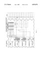
- FIG. 1 is a view of a display showing a matrix of gynecological organs and conditions or complained of symptoms;
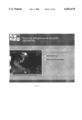
- FIG. 2 is a view of a display showing detailed information about a condition, tubal pregnancy, as it affects an organ, the fallopian tubes;
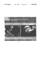
- FIG. 3 is a view of a display showing information about one criteria of a tubal pregnancy, identifying differential diagnosis with probabilities of making a mistake in the shown situation;
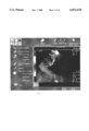
- FIG. 4 is a view of a display showing a side by side comparison of a criteria of a tubal pregnancy and a differential diagnosis with potential for mistake in diagnosis;
- FIG. 5 is a view of a display showing patient data from a sonogram presented in a video display field with organ and condition information surrounding the video display field,
- FIG. 6 is a view of a display showing fetal information arranged by organ system and time-oriented development stage
- FIG. 7 is a representation of the present invention, operating in conjunction with a laparoscope
- FIG. 8 is a representation of a display showing icons superimposed over patient data
- FIG. 9 is a representation of a display showing a side by side comparison of patient data with condition data
- FIG. 10 is a representation of a hierarchical display of the present invention.
- FIG. 11 is a representation showing access routes to the hierarchical display
- FIG. 12 is a view of a display showing access through a glossary of condition and organ information
- FIG. 13 is a view of a display showing a correlation matrix identifying organs and a list of drugs for diagnosis with respect to interactions;
- FIG. 14 is a side by side comparison of a fetus with an anomaly as marked as compared to a normal fetus at the same time period;
- FIG. 15 is an example of a listing of fetal anomalies which leads to the matrix shown in FIG. 1;
- FIG. 16 is the matrix showing classification of iseases according to benign, malignant, borderline, tumorlike and other conditions and list of malignant tumors.
- initial subjective and objective observations are utilized to determine the nature of an initial screening such as a pelvic examination for a gynecological problem (the problem having been suspected for reasons as outlined above).
- the various organs relevant to the condition are screened with an appropriate device such as with a laparoscope and the images obtained are stored and analyzed by a computer by sites of interest for possible conditions.
- the analysis is made, as in the prior art, from answers to a generated questionnaire regarding condition of various "landmarks" of the image (in sophisticated systems, the computer itself can scan and determine the answers with respect to the "landmarks").
- a matrix of possible conditions relative to organs is generated.
- the operator or diagnostician determines, with guidance, the most probable diagnoses and checks definitions of the various conditions which are available at hand with specifics regarding factors which are indicative of the condition.
- a match is determined to have occurred between a basis for condition and the actual image obtained, with a probability above a threshold level, the operator activates the portion of the matrix of the intersection between organ and suspected condition.
- An example of an organ having the suspected condition is displayed for direct visual comparison with the stored image of the patient and a probability of correct diagnosis is assessed.
- the operator is also advised of conditions for error and the various possibilities which can lead to error. Selection of different condition and comparative image continues until the most likely conditions are obtained on a probability basis.
- the device of the present invention provides guidance, in the form of recommendations for the conducting of additional test regimens, e.g., x-rays, to further refine the selected diagnoses for a most likely diagnosis, with caveats with respect to other conditions if the diagnosis is in error.
- additional test regimens e.g., x-rays
- the diagnostician is also guided to explore conditions which may have resulted in a diagnoses but wherein additional conditions may be responsible for such diagnoses being in error.
- the device provides guidance with respect to follow ups (e.g., return visits for additional testing, with conditions which change in a specific manner as indicative of the existence of a condition, within a non-health threatening time period) and a regimen of treatment.
- follow ups e.g., return visits for additional testing, with conditions which change in a specific manner as indicative of the existence of a condition, within a non-health threatening time period
- a report may be generated which includes the diagnosis, both the actual image and corresponding library matched image, and a listing of probable errors with matching error images (i.e. similar but probably erroneous for reasons listed).
- adjunct device of the present invention during the operation of the adjunct device of the present invention it presents a visual display field from an existing diagnosis device surrounded by an additional video display generated by the invention.
- An example of a combined video display is shown in FIG. 5.
- This additional video display contains a two dimensional matrix of information. Along one dimension are representations of anatomical organs of the site being examined, and which are appropriately examined by the diagnosis device. Along the other dimension are representations of conditions (e.g., symptoms being complained of, where such conditions are capable of examination and diagnosis by the diagnosis device.
- the matrix display is identified herein as a "Corematrix,” for correspondence or correlation matrix.
- the organ and condition representations may be placed in any convenient location on the screen, it is desirable to arrange them so they form a matrix, or Corematrix, from which an intersection is readily apparent. This preferred display conserves most of the display for the purpose of viewing the site of interest by any of the available or future methods of diagnosis.
- the organs and the conditions may be displayed in text, or they may be represented by icons, or they may be shown by some combination of text and icons, as shown in FIG. 1.
- the intersection point when activated, relates the conditions to the organ and the adjunct system evaluates the probability of diagnosis with respect to the condition affecting the organ.
- the adjunct device also retrieves from a stored library of images corresponding (as imaged by the same diagnostic device) to the organ and having the condition related by the matrix whereby the actual image can be directly compared to an image with the condition as shown in FIG. 14.
- the adjunct device of the present invention allows for the superimposition of icons over the visual display field, as shown in FIG. 8, where the icons may indicate conditions which affect a represented anatomical organ, or the icons may indicate anatomical organs which may be affected by a represented condition.
- the visual display field may alternate with a grid display of such icons, as is shown in FIG. 1.
- this display may not include all possible organs or conditions, but rather identify an organ subsystem or category of conditions. Selection of a displayed organ subsystem or category of conditions will present an alternate two dimensional matrix containing representations of various anatomical organs or organs within a subsystem along one dimensional axis of the matrix, with representations of various conditions or subsets of a category of conditions along the other dimensional axis.
- the operator or diagnostician may select an icon, superimposed or not, to obtain detailed information about the selected condition as it affects the selected organ. Selection can be accomplished in any way such as with a track ball, mouse or a touch sensitive panel.
- the detailed information preferably includes a visual representation of the condition as it affects the organ in a typical case.
- the detailed information may also include criteria for the condition, presented in textual or graphic format.
- FIG. 2 is an example of detailed information of a tubal pregnancy with a video clip of a sonogram of a typical case, along with text identifying criteria for diagnosis of a tubal pregnancy. Patient data may be shown adjacent to typical condition data for ease of analysis, as shown in FIG. 9.
- the operator or diagnostician may select one or more criteria to obtain additional information.
- additional information preferably includes an alternate visual representation of the condition, showing the chosen criteria in greater detail, as shown in FIG. 3.
- the additional information may also include references to differential diagnoses. Such differential diagnoses are previously categorized and associated with the alternate criteria as selected by the operator.
- the operator would be offered a selection of other conditions that appear similar to the selected condition in the selected organ. These may be listed anywhere on the display, and in any manner, but are preferably listed in thumbnail views on the display below the shown stored image. If any of these conditions are selected, a stored image of the newly selected condition may be displayed in place of the currently shown stored image. And, as above, the operator may be offered a selection of other conditions that appear similar to the selected condition in the selected organ.
- the operator is offered a differential diagnosis.
- the operator may be offered a set of criteria that would also be required to determine that the selected condition is actually present in the selected organ.
- the operator can select any of D1, D2 or D3, and if selected, these would be displayed either in an additional window on the display (not shown) or in place of the stored image
- D1, D2 or D3 would be displayed either in an additional window on the display (not shown) or in place of the stored image
- Selection of the presented differential diagnosis by the operator prompts the adjunct device to display a variation of the initial Corematrix screen, highlighting the selected differential diagnosis as a condition, and identifying the various organs which are affected by the selected diagnosis.
- the operator may select a particular organ affected and repeat the process for the differential diagnosis.
- the laparoscope is provided with a camera that transmits an image of the site of interest to the operator.
- the image is input into a computer system and output on the television-type display which the operator views to observe the site of interest.
- the display also comprises representations of organs that may be encountered in the present laparoscopic procedure along the top, and representations of conditions that may be encountered along the left side.
- the computer superimposes upon the site display a column of the matrix corresponding to the selected organ, and identifying each of the conditions that would apply to this organ. In a preferred embodiment, this is represented by the presence of some symbol at the matrix cross-point between the organ and the corresponding condition.
- the operator may select from the display the condition believed to be present. If this condition is not one that applies to the organ, the selection may then switch to the most appropriate case (based on preset probabilities). If, however, the condition corresponds to the organ, as shown for example, by the presence of a dot, the operator may select that condition. Selection, as described above, may be done by any known means or method.
- the display is changed to simultaneously show the present site of interest in the same field of view with a stored image of the selected organ in the selected condition.
- the stored image is preferably stored on a storage device, such as CD-ROM or optical disks, or a hard disk drive of the computer (see FIG. 7).
- the storage device is not important to the invention in that any type of storage device may be used provided that it can store a plurality of images (still or moving), and can selectively retrieve the images. The operator can now simultaneously view the stored image alongside of the actual site of interest.
- FIG. 10 depicts a representation of the relationship between the hierarchical display and access points, such as the Corematrix display of FIG. 1, or through a glossary function as shown in FIG. 12.
- the operator may access the medical condition information by way of a glossary function, as shown in FIG. 12.
- a glossary function as shown in FIG. 12.
- the database of medical condition information is indexed, according to a set of rules, such as alphabetical by name of the condition. The operator may thereby select a particular medical condition through the glossary. Selection of a medical condition will direct the diagnostic device to the hierarchical page presentation as shown in FIGS. 10 and 11. The operator may navigate through the system as discussed above.
- the invention may also be embodied as a stand-alone system, incorporating data from a plurality of diagnostic tools, where the operator may be first presented with a computer screen identifying a plurality of subsystems to choose from.
- These subsystems may be broken down by various anatomical systems, by disease states, by medical diagnostic device, or by anatomical organ.
- the operator is presented with a two dimensional Corematrix configuration identifying potential data categories for a given medical diagnostic device, shown in FIG. 1 as ultrasonogram categories of a gynecological subsystem.
- the matrix configuration provides representations of various anatomical organs along one dimensional axis of the matrix, with representations of various conditions along the other dimensional axis.
- a three dimensional matrix may also be utilized, where the third dimension may provide for identification of categories of anatomical organs and conditions related to, or detectable by, different medical diagnosis devices.
- the operator's selection of one of the organ representations will result in the invention identifying conditions affecting the represented organ and provide a series of "hot-spots" for the operator to select.
- the invention may thereby be utilized as discussed above.
- the stored library images that are displayed for comparative purposes may be from a variety of data, such as previous images of the same organ in the present patient at an earlier date.
- one or more stored image for each organ-condition combination are used to represent the typical or expected image of the condition in that organ.
- the computer be able to color balance or image process the stored image to match the environment of the laparoscope, or to permit setting of color balance controls or other imaging controls (which may include, e.g., zoom, scale, etc.) by the operator.
- stored images include both still and moving images, to include examination of a site of interest in an organ that normally moves perceptibly, as would be the case with the heart.
- clicking on the appropriate button provides return and activation of the matrix.
Abstract
An adjunct device and system which works in tandem with existing diagnostic imaging tools such as for medical diagnosis to enhance reliability of diagnosis with guidance for appropriate treatment or imaging devices for machinery (or other objects) and guidance for repair. The adjunct device initially analyzes an input image and either automatically or semi-automatically (with input from a grid of organ and anomalies) provides matching images for adjacent viewing and comparison. Where relevant, the device provides weighted possible diagnoses with advisory pathways for treatment or additional testing. The device and system include computer elements with stored medical data appropriate to the diagnostic tool being used and the body part(s) or objects, being diagnosed to provide the appropriate comparative image.
Description
This application claims priority from the filing date of the U.S. Provisional Application 60/039,073 filed on Mar. 14,1997.
This invention relates to devices used as adjuncts to diagnostic imaging systems and devices and particularly to such device adjuncts used for analysis of images of human body organs for the enhancement of correctness of diagnosis thereof by existing diagnostic devices.
The need for conducting a scan such as for medical purposes or for generally determining a particular state of a person or object is triggered by one of three basic factors. In a first instance, the person complains of various symptoms (or an object is similar to other objects which have been exhibiting defects). Secondly the person or object is already being checked for some unrelated reason and an anomaly appears which requires further investigation, or thirdly there is simply an inquiry for the conducting of a screening test to determine (or to rule out) a specific or suspected state or condition (e.g., a prospective parent simply wishing to observe a sonogram of an unborn child).
The basic diagnostic methods of determining the health conditions of a patient, or whether there are anomalies in a patient's medical condition, are primarily:
(a) direct interviews with the patient for the subjective determination of not overtly apparent symptoms (e.g., pain) and conditions, and
(b) the imaging of affected areas of the patient's body for an objective determination (regardless of whether the patient is complaining of a symptom or something was found based on testing for an unrelated condition or state).
The type of imaging most properly utilized for an initial objective determination is usually dictated by the nature of the organ or part of the body exhibiting a specific condition (or which is being tested for determining if a condition exists), or the actual condition which is suspected.
Imaging devices (the term "imaging" used hereinafter includes optical, aural and any other sensory recordable state of an object or patient) primarily include those which permit visual inspection of a site or cavity directly or by use of a lens system for optical enhancement, and devices which permit visual inspection of a site through analog or digital displays or the analysis of images resulting from the use of ultrasound waves (sonograms), magnetic resonance (MRI), computerized tomography (CAT scans), nuclear medicine, x-rays or other imaging technology. Existing specific tools or devices used for imaging include laparoscopes, MRI and ultrasonogram devices, as well as hysteroscopes, arthroscopes, esophagoscopes, bronchoscopes, rectoscopes, laryngoscopes, otoscopes, ophthalmoscopes, colposcopes, microscopes, computed radiography, x-ray imaging, computed tomography, mammography, angiography, gamma camera and nuclear medicine instruments, boreoscopes (used for internal analysis of machinery) and the like, which are all well known diagnostic tools in the art.
Results from use of an imaging devices are usually directly visually (or less often, aurally) analyzed by skilled technicians (e.g., structural analysis of objects such as aircraft for metal fatigue or other possible defects) or medical personnel (i.e., diagnosticians) for determination of abnormalities or the lack thereof, in deciding on a probable basis for a condition or even for the determination that a condition does not exist. However, even with skilled analyzers, anomalies may be slight, obscured, or even not abnormal in the patient being tested since, while generally similar, body parts and organs are rarely sufficiently different in appearance between patients, whereby, unless specifically recognized, diagnostic results may be in error.
To assist in making a diagnosis from an image scanning, oftentime "landmarks" of an object or an organ are designated as being indicative of anomalies and the portion of the image related to the landmark is digitized and computer analyzed with a stored database. This comparison serves to determine whether the portion falls within accepted normal parameters or not, in providing an automatically generated diagnosis. Examples of such systems, with respect to medical diagnosis, are disclosed in U.S. Pat. Nos. 5,235,510 and 5,437,278.
Though the original image remains available for the diagnostician in such computer aided systems, there is little enhancement to the actual analysis by the diagnostician of the image itself except for some indication where and what to look for more closely, in determining the veracity of the digitized analysis. In addition, the results from the digitization are only as valid as the data originally input with acceptance of a diagnosis serving to take away decisional control from the diagnostician. Furthermore, the devices are entirely geared to simple diagnosis based on a comparison between digitized portions of an image and a stored data base. There is however no guidance with respect to appropriate additional tests, probability of correctness of diagnosis and probability of mistake of the diagnosis (with reasons or basis for the possible errors), follow ups, treatment and the like.
It is an object of the present invention to provide a multi-tier diagnostic and treatment, repair and monitoring or follow up advisory system wherein the diagnostician retains comparative control, with a direct comparison of library images (normal and abnormal images closely related to patient type) to the real time image of the patient's organ or body type (or object being imaged), regardless of imaging device utilized.
It is a further object of the present invention to provide the diagnostic and treatment advisory system whereby the system advises the diagnostician, in various stages, of the probability of likely conditions, errors which may result in incorrect diagnoses, the necessary steps or tests for refinement of the diagnoses, treatment relevant to the diagnoses and conditions for re-examination.
Generally the present invention comprises an adjunct for an existing diagnostic and treatment or diagnostic and repair system which is adapted for use by an operator with existing organ and body imaging devices or machinery diagnostic and repair systems and the like. The system comprises:
a) means for providing comparative images for a diagnostician to directly compare on a single display, the image obtained from the patient (or object) and library stored images (from the same type of screening device), corresponding to probable conditions as determined either by the diagnostician or by computerized comparison to the library stored images falling within preset comparison parameters;
b) means for providing weighted probability for a particular diagnosis being relevant to the patient's (or object's) condition, based on library stored general parameters and optionally in further view of the patient's prior medical history (or repair or maintenance history);
c) means for providing weighted probability for a particular mistake being relevant to the patient's (or object's) condition and image based on library stored general parameters and optionally in further view of the patient's prior medical history (or object's prior history) and present image on the screen. (The means also provides a showing of probable errors with respect to a particular selected image).
d) means for providing to the diagnostician, if relevant, which additional test are required to increase the probability of relevance of a probable diagnosis;
e) means for providing to the diagnostician, information regarding treatment or repair protocols for a diagnosis with a probability above a pre-set probability level.
f) means for providing the user or diagnostician case and situation directions for monitoring and follow-up.
These and other objects, features and advantages of the present invention will become more evident from the following discussion and the drawings in which:
FIG. 1 is a view of a display showing a matrix of gynecological organs and conditions or complained of symptoms;
FIG. 2 is a view of a display showing detailed information about a condition, tubal pregnancy, as it affects an organ, the fallopian tubes;
FIG. 3 is a view of a display showing information about one criteria of a tubal pregnancy, identifying differential diagnosis with probabilities of making a mistake in the shown situation;
FIG. 4 is a view of a display showing a side by side comparison of a criteria of a tubal pregnancy and a differential diagnosis with potential for mistake in diagnosis;
FIG. 5 is a view of a display showing patient data from a sonogram presented in a video display field with organ and condition information surrounding the video display field,
FIG. 6 is a view of a display showing fetal information arranged by organ system and time-oriented development stage;
FIG. 7 is a representation of the present invention, operating in conjunction with a laparoscope;
FIG. 8 is a representation of a display showing icons superimposed over patient data;
FIG. 9 is a representation of a display showing a side by side comparison of patient data with condition data;
FIG. 10 is a representation of a hierarchical display of the present invention;
FIG. 11 is a representation showing access routes to the hierarchical display;
FIG. 12 is a view of a display showing access through a glossary of condition and organ information;
FIG. 13 is a view of a display showing a correlation matrix identifying organs and a list of drugs for diagnosis with respect to interactions;
FIG. 14 is a side by side comparison of a fetus with an anomaly as marked as compared to a normal fetus at the same time period;
FIG. 15 is an example of a listing of fetal anomalies which leads to the matrix shown in FIG. 1; and
FIG. 16 is the matrix showing classification of iseases according to benign, malignant, borderline, tumorlike and other conditions and list of malignant tumors.
With the operation of the present invention, initial subjective and objective observations are utilized to determine the nature of an initial screening such as a pelvic examination for a gynecological problem (the problem having been suspected for reasons as outlined above). The various organs relevant to the condition are screened with an appropriate device such as with a laparoscope and the images obtained are stored and analyzed by a computer by sites of interest for possible conditions. The analysis is made, as in the prior art, from answers to a generated questionnaire regarding condition of various "landmarks" of the image (in sophisticated systems, the computer itself can scan and determine the answers with respect to the "landmarks"). A matrix of possible conditions relative to organs is generated. The operator or diagnostician then determines, with guidance, the most probable diagnoses and checks definitions of the various conditions which are available at hand with specifics regarding factors which are indicative of the condition. When a match is determined to have occurred between a basis for condition and the actual image obtained, with a probability above a threshold level, the operator activates the portion of the matrix of the intersection between organ and suspected condition. An example of an organ having the suspected condition is displayed for direct visual comparison with the stored image of the patient and a probability of correct diagnosis is assessed. With the selection of the condition and related image, the operator is also advised of conditions for error and the various possibilities which can lead to error. Selection of different condition and comparative image continues until the most likely conditions are obtained on a probability basis. The device of the present invention provides guidance, in the form of recommendations for the conducting of additional test regimens, e.g., x-rays, to further refine the selected diagnoses for a most likely diagnosis, with caveats with respect to other conditions if the diagnosis is in error. The diagnostician is also guided to explore conditions which may have resulted in a diagnoses but wherein additional conditions may be responsible for such diagnoses being in error.
When the most likely diagnosis has been arrived at with elimination of errors, the device provides guidance with respect to follow ups (e.g., return visits for additional testing, with conditions which change in a specific manner as indicative of the existence of a condition, within a non-health threatening time period) and a regimen of treatment.
After the procedure is completed, a report may be generated which includes the diagnosis, both the actual image and corresponding library matched image, and a listing of probable errors with matching error images (i.e. similar but probably erroneous for reasons listed).
In a preferred embodiment of the invention, during the operation of the adjunct device of the present invention it presents a visual display field from an existing diagnosis device surrounded by an additional video display generated by the invention. An example of a combined video display is shown in FIG. 5.
This additional video display contains a two dimensional matrix of information. Along one dimension are representations of anatomical organs of the site being examined, and which are appropriately examined by the diagnosis device. Along the other dimension are representations of conditions (e.g., symptoms being complained of, where such conditions are capable of examination and diagnosis by the diagnosis device. The matrix display is identified herein as a "Corematrix," for correspondence or correlation matrix.
While the organ and condition representations may be placed in any convenient location on the screen, it is desirable to arrange them so they form a matrix, or Corematrix, from which an intersection is readily apparent. This preferred display conserves most of the display for the purpose of viewing the site of interest by any of the available or future methods of diagnosis. Further, the organs and the conditions may be displayed in text, or they may be represented by icons, or they may be shown by some combination of text and icons, as shown in FIG. 1. The intersection point, when activated, relates the conditions to the organ and the adjunct system evaluates the probability of diagnosis with respect to the condition affecting the organ. The adjunct device also retrieves from a stored library of images corresponding (as imaged by the same diagnostic device) to the organ and having the condition related by the matrix whereby the actual image can be directly compared to an image with the condition as shown in FIG. 14.
The adjunct device of the present invention allows for the superimposition of icons over the visual display field, as shown in FIG. 8, where the icons may indicate conditions which affect a represented anatomical organ, or the icons may indicate anatomical organs which may be affected by a represented condition. Alternatively, the visual display field may alternate with a grid display of such icons, as is shown in FIG. 1.
For ease of use and display, this display may not include all possible organs or conditions, but rather identify an organ subsystem or category of conditions. Selection of a displayed organ subsystem or category of conditions will present an alternate two dimensional matrix containing representations of various anatomical organs or organs within a subsystem along one dimensional axis of the matrix, with representations of various conditions or subsets of a category of conditions along the other dimensional axis. The operator or diagnostician may select an icon, superimposed or not, to obtain detailed information about the selected condition as it affects the selected organ. Selection can be accomplished in any way such as with a track ball, mouse or a touch sensitive panel.
The detailed information preferably includes a visual representation of the condition as it affects the organ in a typical case. The detailed information may also include criteria for the condition, presented in textual or graphic format. FIG. 2 is an example of detailed information of a tubal pregnancy with a video clip of a sonogram of a typical case, along with text identifying criteria for diagnosis of a tubal pregnancy. Patient data may be shown adjacent to typical condition data for ease of analysis, as shown in FIG. 9.
The operator or diagnostician may select one or more criteria to obtain additional information. Such additional information preferably includes an alternate visual representation of the condition, showing the chosen criteria in greater detail, as shown in FIG. 3. The additional information may also include references to differential diagnoses. Such differential diagnoses are previously categorized and associated with the alternate criteria as selected by the operator.
In a preferred embodiment, the operator would be offered a selection of other conditions that appear similar to the selected condition in the selected organ. These may be listed anywhere on the display, and in any manner, but are preferably listed in thumbnail views on the display below the shown stored image. If any of these conditions are selected, a stored image of the newly selected condition may be displayed in place of the currently shown stored image. And, as above, the operator may be offered a selection of other conditions that appear similar to the selected condition in the selected organ.
Also in a preferred embodiment, the operator is offered a differential diagnosis. For example, the operator may be offered a set of criteria that would also be required to determine that the selected condition is actually present in the selected organ. In FIG. 9, the operator can select any of D1, D2 or D3, and if selected, these would be displayed either in an additional window on the display (not shown) or in place of the stored image For all available or future diagnostic tools it is preferred not to obscure the real-time or post-processing operator's view of the site of interest.
Selection of the presented differential diagnosis by the operator prompts the adjunct device to display a variation of the initial Corematrix screen, highlighting the selected differential diagnosis as a condition, and identifying the various organs which are affected by the selected diagnosis. The operator may select a particular organ affected and repeat the process for the differential diagnosis.
With reference to FIG. 7, and the shown utilization of a laparoscopic for imaging, the laparoscope is provided with a camera that transmits an image of the site of interest to the operator. The image is input into a computer system and output on the television-type display which the operator views to observe the site of interest. The display also comprises representations of organs that may be encountered in the present laparoscopic procedure along the top, and representations of conditions that may be encountered along the left side. When the operator points the laparoscope camera at a particular site of interest, for example, the fallopian tubes, such organ can be selected from the display.
With reference to FIG. 8, once the organ is selected, (organ 2 in FIG. 8) the computer superimposes upon the site display a column of the matrix corresponding to the selected organ, and identifying each of the conditions that would apply to this organ. In a preferred embodiment, this is represented by the presence of some symbol at the matrix cross-point between the organ and the corresponding condition. When the operator detects a condition in the organ, the operator may select from the display the condition believed to be present. If this condition is not one that applies to the organ, the selection may then switch to the most appropriate case (based on preset probabilities). If, however, the condition corresponds to the organ, as shown for example, by the presence of a dot, the operator may select that condition. Selection, as described above, may be done by any known means or method.
With respect to FIG. 9, once both an organ and a condition are selected, the display is changed to simultaneously show the present site of interest in the same field of view with a stored image of the selected organ in the selected condition. The stored image is preferably stored on a storage device, such as CD-ROM or optical disks, or a hard disk drive of the computer (see FIG. 7). The storage device, however, is not important to the invention in that any type of storage device may be used provided that it can store a plurality of images (still or moving), and can selectively retrieve the images. The operator can now simultaneously view the stored image alongside of the actual site of interest.
The various functions of the invention may be accessed through a master, or hierarchical display, as shown in FIG. 10. The hierarchical display provides an overview of the relations of the various diagnosis points of the invention, whereby the operator may access any given point without having to follow through a prescribed order of steps. FIG. 11 depicts a representation of the relationship between the hierarchical display and access points, such as the Corematrix display of FIG. 1, or through a glossary function as shown in FIG. 12.
The operator may access the medical condition information by way of a glossary function, as shown in FIG. 12. Using the glossary, the database of medical condition information is indexed, according to a set of rules, such as alphabetical by name of the condition. The operator may thereby select a particular medical condition through the glossary. Selection of a medical condition will direct the diagnostic device to the hierarchical page presentation as shown in FIGS. 10 and 11. The operator may navigate through the system as discussed above.
The invention may also be embodied as a stand-alone system, incorporating data from a plurality of diagnostic tools, where the operator may be first presented with a computer screen identifying a plurality of subsystems to choose from. These subsystems may be broken down by various anatomical systems, by disease states, by medical diagnostic device, or by anatomical organ.
Once a subsystem is selected, the operator is presented with a two dimensional Corematrix configuration identifying potential data categories for a given medical diagnostic device, shown in FIG. 1 as ultrasonogram categories of a gynecological subsystem. In a preferred embodiment, the matrix configuration provides representations of various anatomical organs along one dimensional axis of the matrix, with representations of various conditions along the other dimensional axis. A three dimensional matrix may also be utilized, where the third dimension may provide for identification of categories of anatomical organs and conditions related to, or detectable by, different medical diagnosis devices.
The operator's selection of one of the organ representations will result in the invention identifying conditions affecting the represented organ and provide a series of "hot-spots" for the operator to select. The invention may thereby be utilized as discussed above.
Although the above description is described with reference to a laparoscope, it would be similar for any other tool disclosed above. Where the tool does not have its own display, numerous options are available. First, it is possible to implement the above-described invention within the field of view of, for example, a microscope or otoscope where the outer perimeter of a generally circular view could be used to present organs and conditions (rather than the top and side of a rectangular screen). Alternatively, it is possible to implement the above-described invention within the field of view of a purely mechanical device, as a speculum, by fastening a display screen to the device which provides for a simultaneous viewing of the site of interest and display of stored images.
In lieu of the side by side analysis it is also within the ambit of the invention to accept an output from an existing diagnostic tool and superimpose the stored image and the selection images upon it, or to feed the stored image and the selection images to the existing tool and have the existing display provide all of the necessary information in the operator's field of view.
The stored library images that are displayed for comparative purposes may be from a variety of data, such as previous images of the same organ in the present patient at an earlier date. In another preferred embodiment one or more stored image for each organ-condition combination are used to represent the typical or expected image of the condition in that organ.
It is also preferred that the computer be able to color balance or image process the stored image to match the environment of the laparoscope, or to permit setting of color balance controls or other imaging controls (which may include, e.g., zoom, scale, etc.) by the operator.
It is understood that stored images include both still and moving images, to include examination of a site of interest in an organ that normally moves perceptibly, as would be the case with the heart.
Though this description has been given with an organ bias, it is also possible to first select a condition, and then select the organ affected. In other words, it is possible to carry out the invention with a condition bias. Alternatively, information about drugs or time periods may be utilized, as shown in FIGS. 6 and 13.
With respect to FIGS. 15 and 16, clicking on the appropriate button provides return and activation of the matrix.
It is possible, without departing from the spirit of the invention to supply substantial additional information to the operator's field of view. For example, in some applications, it would be preferred to include additional diagnostic information, drug interactions, etc.
It is understood that the above description and drawings are merely exemplary of the present invention and that changes in matrixes, organs being scanned or screened and conditions may be made without departing from the scope of the present invention. In addition, changes may be made to the manner and type of information and images being provided as well as the subject matter being examined. Thus, there is, in addition to application to medical diagnoses, similar application to any field requiring a diagnosis such as instrument examination of machines, and objects such as airplanes and the like, subject to varying conditions (e.g., metal fatigue, rust, leaks, etc. ) and requiring diagnosis and comparison to existing established criteria for defects and conditions. These and other changes and modifications are possible without departing from the scope of the present invention as defined in the following claims.
Claims (4)
1. An adjunct device and system for use with an existing diagnostic object or body imaging device, said adjunct device comprising:
a) means for providing comparative images to directly compare on a single display an image of an object or body obtained from the imaging device and one or more library stored images from the same type of imaging device of a corresponding object or body having probable conditions as the object or body being imaged, the probable conditions being determined either by the user or by a computerized comparison of the image to the library stored images falling within preset comparison parameters; whereby a probable diagnosis of an actual condition of the object or body can be formulated by a matching comparison;
b) means for providing a weighted probability for a particular diagnosis being relevant to said condition, said weighted probability being based on library stored general parameters and optionally in further view of the prior history of the body or object;
c) means for providing a weighted probability for a particular mistake being relevant to the condition and image based on library stored general parameters and optionally in further view of the prior history of the body or object and present image on the screen;
d) means for providing to the user, if relevant, which additional test or tests are required to increase the probability of relevance of a probable diagnosis;
e) means for providing to the user a case and situation oriented need for follow-up and/or monitoring of conditions; and
f) means for providing to the user, information regarding treatment or repair protocols, as relevant to the body or object, for a diagnosis with a probability above a pre-set probability level.
2. The adjunct device of claim 1 wherein the condition is a medical condition and the imaging device is adapted for imaging of human body parts or organs.
3. The adjunct device of claim 1 wherein the condition is a mechanical condition of a machine or device.
4. The adjunct device of claim 1 wherein the means for providing weighted probability for a particular mistake being relevant also provides a showing of probable errors with respect to a particular image selected from the library stored images.
Priority Applications (1)
| Application Number | Priority Date | Filing Date | Title |
|---|---|---|---|
| US09/042,269 US6032678A (en) | 1997-03-14 | 1998-03-13 | Adjunct to diagnostic imaging systems for analysis of images of an object or a body part or organ |
Applications Claiming Priority (2)
| Application Number | Priority Date | Filing Date | Title |
|---|---|---|---|
| US3907397P | 1997-03-14 | 1997-03-14 | |
| US09/042,269 US6032678A (en) | 1997-03-14 | 1998-03-13 | Adjunct to diagnostic imaging systems for analysis of images of an object or a body part or organ |
Publications (1)
| Publication Number | Publication Date |
|---|---|
| US6032678A true US6032678A (en) | 2000-03-07 |
Family
ID=26715794
Family Applications (1)
| Application Number | Title | Priority Date | Filing Date |
|---|---|---|---|
| US09/042,269 Expired - Fee Related US6032678A (en) | 1997-03-14 | 1998-03-13 | Adjunct to diagnostic imaging systems for analysis of images of an object or a body part or organ |
Country Status (1)
| Country | Link |
|---|---|
| US (1) | US6032678A (en) |
Cited By (94)
| Publication number | Priority date | Publication date | Assignee | Title |
|---|---|---|---|---|
| WO2000025191A2 (en) * | 1998-10-26 | 2000-05-04 | Visionary Medical, Inc. | Portable data collection device |
| US6327490B1 (en) | 1998-02-27 | 2001-12-04 | Varian Medical Systems, Inc. | Brachytherapy system for prostate cancer treatment with computer implemented systems and processes to facilitate pre-implantation planning and post-implantation evaluations with storage of multiple plan variations for a single patient |
| US6349330B1 (en) * | 1997-11-07 | 2002-02-19 | Eigden Video | Method and appparatus for generating a compact post-diagnostic case record for browsing and diagnostic viewing |
| US20020028010A1 (en) * | 2000-09-05 | 2002-03-07 | Fuji Photo Film Co., Ltd. | Method and apparatus for outputting optical tomographic image diagnostic data |
| US6360116B1 (en) | 1998-02-27 | 2002-03-19 | Varian Medical Systems, Inc. | Brachytherapy system for prostate cancer treatment with computer implemented systems and processes to facilitate pre-operative planning and post-operative evaluations |
| WO2002039891A1 (en) * | 2000-10-06 | 2002-05-23 | Ultratouch Corporation | A dynamic health metric reporting method and system |
| US6400837B2 (en) | 1997-10-24 | 2002-06-04 | Ultratouch Corporation | Location head for an apparatus for detecting very small breast anomalies |
| US20020068857A1 (en) * | 2000-02-14 | 2002-06-06 | Iliff Edwin C. | Automated diagnostic system and method including reuse of diagnostic objects |
| US6453058B1 (en) * | 1999-06-07 | 2002-09-17 | Siemens Corporate Research, Inc. | Computer-assisted diagnosis method using correspondence checking and change detection of salient features in digital images |
| US6459920B1 (en) * | 1999-03-04 | 2002-10-01 | Zila, Inc. | Method for detecting and diagnosing epithelial cancer |
| US6488627B1 (en) * | 1999-11-26 | 2002-12-03 | Medison Co., Ltd. | Ultrasonic image searching apparatus and ultrasonic image transmission and reception system adopting the same |
| US20030036686A1 (en) * | 1997-03-13 | 2003-02-20 | Iliff Edwin C. | Disease management system and method including analysis of disease specific changes |
| WO2003046810A1 (en) * | 2001-11-21 | 2003-06-05 | Wake Forest University Health Sciences | Image reporting method and system |
| US6597938B2 (en) * | 2001-08-16 | 2003-07-22 | Koninklijke Philips Electronics, N.V. | System for assistance of parameter determination and diagnosis in MRI dynamic uptake studies |
| US20030163299A1 (en) * | 1993-12-29 | 2003-08-28 | Iliff Edwin C. | Computerized medical diagnostic and treatment advice system |
| US20030229278A1 (en) * | 2002-06-06 | 2003-12-11 | Usha Sinha | Method and system for knowledge extraction from image data |
| US6675039B2 (en) * | 2001-08-31 | 2004-01-06 | Ge Medical Systems Global Technology Company, Llc | Computed tomography scan protocol |
| EP1380003A2 (en) * | 2001-03-09 | 2004-01-14 | Shraga Rottem | Method and system for enhancing the quality of device images |
| US20040030672A1 (en) * | 2001-08-01 | 2004-02-12 | Garwin Jeffrey L | Dynamic health metric reporting method and system |
| US20040059200A1 (en) * | 1996-07-12 | 2004-03-25 | Iliff Edwin C. | Computerized medical diagnostic system utilizing list-based processing |
| US20040101177A1 (en) * | 2002-11-21 | 2004-05-27 | Siemens Aktiengesellschaft | Method and system for retrieving a medical picture |
| US20040122307A1 (en) * | 2001-11-21 | 2004-06-24 | Shraga Rottem | Method and system for enhancing the quality of device images |
| US20040133083A1 (en) * | 2002-11-13 | 2004-07-08 | Siemens Corporate Research Inc. | System and method for real-time feature sensitivity analysis based on contextual information |
| US20040147840A1 (en) * | 2002-11-08 | 2004-07-29 | Bhavani Duggirala | Computer aided diagnostic assistance for medical imaging |
| US20040193036A1 (en) * | 2003-03-12 | 2004-09-30 | Zhou Xiang Sean | System and method for performing probabilistic classification and decision support using multidimensional medical image databases |
| US20040225223A1 (en) * | 2003-04-25 | 2004-11-11 | Olympus Corporation | Image display apparatus, image display method, and computer program |
| US20050010444A1 (en) * | 2003-06-06 | 2005-01-13 | Iliff Edwin C. | System and method for assisting medical diagnosis using an anatomic system and cause matrix |
| US20050020886A1 (en) * | 2003-07-23 | 2005-01-27 | Ge Medical Systems Information Technologies, Inc. | Monitoring system and method using rules |
| US20050038311A1 (en) * | 2002-09-11 | 2005-02-17 | Siemens Aktiengesellschaft | Device to make expert knowledge accessible for the operation of medical examination devices |
| US20050063576A1 (en) * | 2003-07-29 | 2005-03-24 | Krantz David A. | System and method for utilizing shape analysis to assess fetal abnormality |
| US20050113680A1 (en) * | 2003-10-29 | 2005-05-26 | Yoshihiro Ikeda | Cerebral ischemia diagnosis assisting apparatus, X-ray computer tomography apparatus, and apparatus for aiding diagnosis and treatment of acute cerebral infarct |
| US20050119570A1 (en) * | 2003-12-01 | 2005-06-02 | Stephen Lewis | Ultrasonic image and visualization aid |
| US20050147284A1 (en) * | 1999-08-09 | 2005-07-07 | Vining David J. | Image reporting method and system |
| US20060023966A1 (en) * | 1994-10-27 | 2006-02-02 | Vining David J | Method and system for producing interactive, three-dimensional renderings of selected body organs having hollow lumens to enable simulated movement through the lumen |
| US20060034521A1 (en) * | 2004-07-16 | 2006-02-16 | Sectra Imtec Ab | Computer program product and method for analysis of medical image data in a medical imaging system |
| US20060123002A1 (en) * | 2004-12-08 | 2006-06-08 | Siemens Aktiengesellschaft | Operating method for a computer and corresponding devices |
| WO2006058738A1 (en) * | 2004-12-02 | 2006-06-08 | Lieven Van Hoe | Image evaluation system, methods and database |
| US20060135859A1 (en) * | 2004-10-22 | 2006-06-22 | Iliff Edwin C | Matrix interface for medical diagnostic and treatment advice system and method |
| US20070136090A1 (en) * | 2005-12-12 | 2007-06-14 | General Electric Company | System and method for macro-enhanced clinical workflow |
| US20070165049A1 (en) * | 2005-10-14 | 2007-07-19 | General Electric Company | Configurable system and method for results review |
| US20070172100A1 (en) * | 2006-01-20 | 2007-07-26 | Sakura Finetek U.S.A., Inc. | Automated system of processing biological specimens and method |
| US7306560B2 (en) | 1993-12-29 | 2007-12-11 | Clinical Decision Support, Llc | Computerized medical diagnostic and treatment advice system including network access |
| WO2007147059A2 (en) * | 2006-06-15 | 2007-12-21 | Revolutions Medical Corporation | System for and method of performing a medical evaluation |
| US20080030192A1 (en) * | 2004-07-21 | 2008-02-07 | Hitachi Medical Corporation | Tomographic System |
| US20080037876A1 (en) * | 1999-08-09 | 2008-02-14 | Michael Galperin | Object based image retrieval |
| US7337121B1 (en) * | 1999-03-30 | 2008-02-26 | Iso Claims Services, Inc. | Claim assessment model |
| US20080104116A1 (en) * | 2004-12-02 | 2008-05-01 | Lieven Van Hoe | Image Evaluation System, Methods and Database |
| US20080140708A1 (en) * | 2002-01-31 | 2008-06-12 | Oren Fuerst | System and method for providing a computer aided medical diagnostic over a network |
| US20080208048A1 (en) * | 2007-02-27 | 2008-08-28 | Kabushiki Kaisha Toshiba | Ultrasonic diagnosis support system, ultrasonic imaging apparatus, and ultrasonic diagnosis support method |
| US20080228526A1 (en) * | 2006-09-19 | 2008-09-18 | Christopher Brian Locke | System and method for managing history of patient and wound therapy treatment |
| US20080286734A1 (en) * | 2005-07-20 | 2008-11-20 | Shraga Rottem | Method and Device For Image Quality Accreditation, Hands on Cme, and For Control and Analysis of Accreditation at the Enterprise Level |
| US20090037836A1 (en) * | 2007-07-31 | 2009-02-05 | Caterpillar Inc. | Interaction matrix creation tool |
| US20090082637A1 (en) * | 2007-09-21 | 2009-03-26 | Michael Galperin | Multi-modality fusion classifier with integrated non-imaging factors |
| US20090094053A1 (en) * | 2007-10-09 | 2009-04-09 | Edward Jung | Diagnosis through graphical representation of patient characteristics |
| US7780595B2 (en) | 2003-05-15 | 2010-08-24 | Clinical Decision Support, Llc | Panel diagnostic method and system |
| US20100261979A1 (en) * | 2006-09-22 | 2010-10-14 | Masimo Corporation | Modular patient monitor |
| US20100265251A1 (en) * | 1997-02-25 | 2010-10-21 | Vining David J | Virtual Endoscopy with Improved Image Segmentation and Lesion Detection |
| US20100312070A1 (en) * | 2003-12-02 | 2010-12-09 | Shraga Rottem | Artificial Intelligence and Device for Diagnosis, Screening, Prevention and Treatment of Materno-Fetal Conditions |
| AU2008200088B2 (en) * | 2003-04-25 | 2011-03-24 | Olympus Corporation | Image Display Apparatus, Image Display Method and Image Display Program |
| US20110119092A1 (en) * | 2007-08-07 | 2011-05-19 | Szela Jr Erwin G | Electronic health management system |
| US20110166879A1 (en) * | 2008-09-26 | 2011-07-07 | Koninklijke Philips Electronics N.V. | System and method for fusing clinical and image features for computer-aided diagnosis |
| USRE43433E1 (en) | 1993-12-29 | 2012-05-29 | Clinical Decision Support, Llc | Computerized medical diagnostic and treatment advice system |
| US20120174023A1 (en) * | 2011-01-04 | 2012-07-05 | International Business Machines Corporation | Single page multi-tier catalog browser |
| WO2012131524A2 (en) | 2011-03-29 | 2012-10-04 | Koninklijke Philips Electronics N.V. | Image acquisition and/or image related parameter recommender |
| US20120290957A1 (en) * | 2011-05-12 | 2012-11-15 | Jonathan Chernilo | User interface for medical diagnosis |
| US20130253940A1 (en) * | 2012-03-20 | 2013-09-26 | Zilla Collections Llc | System and method for diagnosis involving crowdsourcing |
| US8620044B2 (en) | 2003-04-25 | 2013-12-31 | Olympus Corporation | Image display apparatus, image display method, and computer program |
| US8724871B1 (en) | 2011-12-14 | 2014-05-13 | Atti International Services Company, Inc. | Method and system for identifying anomalies in medical images |
| US8840549B2 (en) | 2006-09-22 | 2014-09-23 | Masimo Corporation | Modular patient monitor |
| US9113831B2 (en) | 2002-03-25 | 2015-08-25 | Masimo Corporation | Physiological measurement communications adapter |
| WO2015128429A1 (en) * | 2014-02-26 | 2015-09-03 | Grain Ip | Method and system for assisting determination of a medical condition |
| US9153112B1 (en) | 2009-12-21 | 2015-10-06 | Masimo Corporation | Modular patient monitor |
| US9401021B1 (en) | 2011-12-14 | 2016-07-26 | Atti International Services Company, Inc. | Method and system for identifying anomalies in medical images especially those including body parts having symmetrical properties |
| US9436645B2 (en) | 2011-10-13 | 2016-09-06 | Masimo Corporation | Medical monitoring hub |
| USD788312S1 (en) | 2012-02-09 | 2017-05-30 | Masimo Corporation | Wireless patient monitoring device |
| US9779504B1 (en) | 2011-12-14 | 2017-10-03 | Atti International Services Company, Inc. | Method and system for identifying anomalies in medical images especially those including one of a pair of symmetric body parts |
| US20180004899A1 (en) * | 2016-07-01 | 2018-01-04 | Topcon Corporation | Method and system for medical support |
| US9943269B2 (en) | 2011-10-13 | 2018-04-17 | Masimo Corporation | System for displaying medical monitoring data |
| US10226187B2 (en) | 2015-08-31 | 2019-03-12 | Masimo Corporation | Patient-worn wireless physiological sensor |
| US10255997B2 (en) | 2016-07-12 | 2019-04-09 | Mindshare Medical, Inc. | Medical analytics system |
| US10307111B2 (en) | 2012-02-09 | 2019-06-04 | Masimo Corporation | Patient position detection system |
| US10430507B2 (en) * | 2013-07-17 | 2019-10-01 | Canon Kabushiki Kaisha | Report creating support apparatus, method for the same, and computer-readable storage medium |
| US10617302B2 (en) | 2016-07-07 | 2020-04-14 | Masimo Corporation | Wearable pulse oximeter and respiration monitor |
| US10765592B2 (en) | 2006-09-19 | 2020-09-08 | Kci Licensing, Inc. | System and method for determining a fill status of a canister of fluid in a reduced pressure treatment system |
| US10806835B2 (en) | 2006-09-19 | 2020-10-20 | Kci Licensing, Inc. | Reduced pressure treatment system having blockage clearing and dual-zone pressure protection capabilities |
| US10825568B2 (en) | 2013-10-11 | 2020-11-03 | Masimo Corporation | Alarm notification system |
| US10833983B2 (en) | 2012-09-20 | 2020-11-10 | Masimo Corporation | Intelligent medical escalation process |
| CN112288683A (en) * | 2020-06-30 | 2021-01-29 | 深圳市智影医疗科技有限公司 | Pulmonary tuberculosis judgment device and method based on multi-mode fusion |
| US11076777B2 (en) | 2016-10-13 | 2021-08-03 | Masimo Corporation | Systems and methods for monitoring orientation to reduce pressure ulcer formation |
| US11109818B2 (en) | 2018-04-19 | 2021-09-07 | Masimo Corporation | Mobile patient alarm display |
| US11229732B2 (en) | 2006-09-19 | 2022-01-25 | Kci Licensing, Inc. | System and method for locating fluid leaks at a drape of a reduced pressure delivery system |
| USD974193S1 (en) | 2020-07-27 | 2023-01-03 | Masimo Corporation | Wearable temperature measurement device |
| USD980091S1 (en) | 2020-07-27 | 2023-03-07 | Masimo Corporation | Wearable temperature measurement device |
| USD1000975S1 (en) | 2021-09-22 | 2023-10-10 | Masimo Corporation | Wearable temperature measurement device |
Citations (4)
| Publication number | Priority date | Publication date | Assignee | Title |
|---|---|---|---|---|
| US5235510A (en) * | 1990-11-22 | 1993-08-10 | Kabushiki Kaisha Toshiba | Computer-aided diagnosis system for medical use |
| US5251131A (en) * | 1991-07-31 | 1993-10-05 | Thinking Machines Corporation | Classification of data records by comparison of records to a training database using probability weights |
| US5799100A (en) * | 1996-06-03 | 1998-08-25 | University Of South Florida | Computer-assisted method and apparatus for analysis of x-ray images using wavelet transforms |
| US5850465A (en) * | 1989-06-26 | 1998-12-15 | Fuji Photo Film Co., Ltd. | Abnormnal pattern detecting or judging apparatus, circular pattern judging apparatus, and image finding apparatus |
-
1998
- 1998-03-13 US US09/042,269 patent/US6032678A/en not_active Expired - Fee Related
Patent Citations (4)
| Publication number | Priority date | Publication date | Assignee | Title |
|---|---|---|---|---|
| US5850465A (en) * | 1989-06-26 | 1998-12-15 | Fuji Photo Film Co., Ltd. | Abnormnal pattern detecting or judging apparatus, circular pattern judging apparatus, and image finding apparatus |
| US5235510A (en) * | 1990-11-22 | 1993-08-10 | Kabushiki Kaisha Toshiba | Computer-aided diagnosis system for medical use |
| US5251131A (en) * | 1991-07-31 | 1993-10-05 | Thinking Machines Corporation | Classification of data records by comparison of records to a training database using probability weights |
| US5799100A (en) * | 1996-06-03 | 1998-08-25 | University Of South Florida | Computer-assisted method and apparatus for analysis of x-ray images using wavelet transforms |
Cited By (213)
| Publication number | Priority date | Publication date | Assignee | Title |
|---|---|---|---|---|
| USRE43433E1 (en) | 1993-12-29 | 2012-05-29 | Clinical Decision Support, Llc | Computerized medical diagnostic and treatment advice system |
| US9005119B2 (en) | 1993-12-29 | 2015-04-14 | Clinical Decision Support, Llc | Computerized medical diagnostic and treatment advice system including network access |
| US8337409B2 (en) | 1993-12-29 | 2012-12-25 | Clinical Decision Support Llc | Computerized medical diagnostic system utilizing list-based processing |
| USRE43548E1 (en) | 1993-12-29 | 2012-07-24 | Clinical Decision Support, Llc | Computerized medical diagnostic and treatment advice system |
| US20030163299A1 (en) * | 1993-12-29 | 2003-08-28 | Iliff Edwin C. | Computerized medical diagnostic and treatment advice system |
| US7306560B2 (en) | 1993-12-29 | 2007-12-11 | Clinical Decision Support, Llc | Computerized medical diagnostic and treatment advice system including network access |
| US7300402B2 (en) | 1993-12-29 | 2007-11-27 | Clinical Decision Support, Llc | Computerized medical diagnostic and treatment advice system |
| US7297111B2 (en) | 1993-12-29 | 2007-11-20 | Clinical Decision Support, Llc | Computerized medical diagnostic and treatment advice system |
| US8015138B2 (en) | 1993-12-29 | 2011-09-06 | Clinical Decision Support, Llc | Computerized medical self-diagnostic and treatment advice system |
| US20100328305A1 (en) * | 1994-10-27 | 2010-12-30 | Vining David J | Method and System for Producing Interactive, Three-Dimensional Renderings of Selected Body Organs Having Hollow Lumens to Enable Simulated Movement Through the Lumen |
| US8145292B2 (en) | 1994-10-27 | 2012-03-27 | Wake Forest University Health Sciences | Method and system for producing interactive, three-dimensional renderings of selected body organs having hollow lumens to enable simulated movement through the lumen |
| US20060023966A1 (en) * | 1994-10-27 | 2006-02-02 | Vining David J | Method and system for producing interactive, three-dimensional renderings of selected body organs having hollow lumens to enable simulated movement through the lumen |
| US7792565B2 (en) | 1994-10-27 | 2010-09-07 | Wake Forest University Health Sciences | Method and system for producing interactive, three-dimensional renderings of selected body organs having hollow lumens to enable simulated movement through the lumen |
| US7344496B2 (en) | 1996-07-12 | 2008-03-18 | Clinical Decision Support, Llc | Computerized medical diagnostic system utilizing list-based processing |
| US20040059200A1 (en) * | 1996-07-12 | 2004-03-25 | Iliff Edwin C. | Computerized medical diagnostic system utilizing list-based processing |
| US20100265251A1 (en) * | 1997-02-25 | 2010-10-21 | Vining David J | Virtual Endoscopy with Improved Image Segmentation and Lesion Detection |
| US8682045B2 (en) | 1997-02-25 | 2014-03-25 | Wake Forest University Health Sciences | Virtual endoscopy with improved image segmentation and lesion detection |
| US8392217B2 (en) | 1997-03-13 | 2013-03-05 | Clinical Decision Support, Llc | Disease management system and method including preview mode |
| US8727976B2 (en) | 1997-03-13 | 2014-05-20 | Clinical Decision Support, Llc | Disease management system operating on a network |
| US20080052120A1 (en) * | 1997-03-13 | 2008-02-28 | Clinical Decision Support, Llc | Disease management system and health assessment method |
| US20080052130A1 (en) * | 1997-03-13 | 2008-02-28 | Clinical Decision Support, Llc. | Disease management system and method including therapy optimization |
| US20080052132A1 (en) * | 1997-03-13 | 2008-02-28 | Clinical Decision Support, Llc | Disease management system and method including therapeutic alterations permission level |
| US20080051641A1 (en) * | 1997-03-13 | 2008-02-28 | Clinical Decision Support , Llc | Disease management system including a no response method |
| US7769600B2 (en) | 1997-03-13 | 2010-08-03 | Clinical Decision Support | Disease management system and method |
| US8066636B2 (en) | 1997-03-13 | 2011-11-29 | Clinical Decision Support, Llc | Disease management system and method including pain code |
| US7297108B2 (en) | 1997-03-13 | 2007-11-20 | Clinical Decision Support, Llc | Disease management system and method including analysis of disease specific changes |
| US7993267B2 (en) | 1997-03-13 | 2011-08-09 | Clinical Decision Support, Llc | Disease management system including a no response method |
| US8740790B2 (en) | 1997-03-13 | 2014-06-03 | Clinical Decision Support, Llc | Disease management system and method |
| US8628470B1 (en) | 1997-03-13 | 2014-01-14 | Clinical Decision Support, Llc | Disease management system and method including medication therapy self-management |
| US8630875B2 (en) | 1997-03-13 | 2014-01-14 | Clinical Decision Support, Llc | Disease management system and health assessment method |
| US8727979B2 (en) | 1997-03-13 | 2014-05-20 | Clinical Decision Support, Llc | Disease management system and method including significant symptom filtering |
| US8060378B2 (en) | 1997-03-13 | 2011-11-15 | Clinical Decision Support, Llc | Disease management system and method including question version |
| US20030036686A1 (en) * | 1997-03-13 | 2003-02-20 | Iliff Edwin C. | Disease management system and method including analysis of disease specific changes |
| US8682694B2 (en) | 1997-03-13 | 2014-03-25 | Clinical Decision Support, Llc | Disease management system and method including permission database |
| US8663104B2 (en) | 1997-03-13 | 2014-03-04 | Clinical Decision Support, Llc | Disease management system and method including therapeutic alterations permission level |
| US6400837B2 (en) | 1997-10-24 | 2002-06-04 | Ultratouch Corporation | Location head for an apparatus for detecting very small breast anomalies |
| US6507663B2 (en) | 1997-10-24 | 2003-01-14 | Ultratouch Corporation | Method and apparatus for detecting very small breast anomalies |
| US6349330B1 (en) * | 1997-11-07 | 2002-02-19 | Eigden Video | Method and appparatus for generating a compact post-diagnostic case record for browsing and diagnostic viewing |
| US6327490B1 (en) | 1998-02-27 | 2001-12-04 | Varian Medical Systems, Inc. | Brachytherapy system for prostate cancer treatment with computer implemented systems and processes to facilitate pre-implantation planning and post-implantation evaluations with storage of multiple plan variations for a single patient |
| US6360116B1 (en) | 1998-02-27 | 2002-03-19 | Varian Medical Systems, Inc. | Brachytherapy system for prostate cancer treatment with computer implemented systems and processes to facilitate pre-operative planning and post-operative evaluations |
| US6539247B2 (en) | 1998-02-27 | 2003-03-25 | Varian Medical Systems, Inc. | Brachytherapy system for prostate cancer treatment with computer implemented systems and processes to facilitate pre-implantation planning and post-implantation evaluations with storage of multiple plan variations for a single patient |
| WO2000025191A3 (en) * | 1998-10-26 | 2000-10-12 | Visionary Medical Inc | Portable data collection device |
| WO2000025191A2 (en) * | 1998-10-26 | 2000-05-04 | Visionary Medical, Inc. | Portable data collection device |
| US6319199B1 (en) * | 1998-10-26 | 2001-11-20 | David M. Sheehan | Portable data collection device |
| US6459920B1 (en) * | 1999-03-04 | 2002-10-01 | Zila, Inc. | Method for detecting and diagnosing epithelial cancer |
| US7337121B1 (en) * | 1999-03-30 | 2008-02-26 | Iso Claims Services, Inc. | Claim assessment model |
| US6453058B1 (en) * | 1999-06-07 | 2002-09-17 | Siemens Corporate Research, Inc. | Computer-assisted diagnosis method using correspondence checking and change detection of salient features in digital images |
| US7835560B2 (en) | 1999-08-09 | 2010-11-16 | Wake Forest University Health Sciences | Image reporting method and system |
| US20080152206A1 (en) * | 1999-08-09 | 2008-06-26 | Vining David J | Image reporting method and system |
| US20050147284A1 (en) * | 1999-08-09 | 2005-07-07 | Vining David J. | Image reporting method and system |
| US6785410B2 (en) * | 1999-08-09 | 2004-08-31 | Wake Forest University Health Sciences | Image reporting method and system |
| US8320651B2 (en) | 1999-08-09 | 2012-11-27 | Wake Forest University Health Sciences | Image reporting method and system |
| US7995823B2 (en) | 1999-08-09 | 2011-08-09 | Wake Forest University Health Sciences | Image reporting method and system |
| US20110135174A1 (en) * | 1999-08-09 | 2011-06-09 | Vining David J | Image reporting method and system |
| US7289651B2 (en) | 1999-08-09 | 2007-10-30 | Wake Forest University Health Science | Image reporting method and system |
| US20080037876A1 (en) * | 1999-08-09 | 2008-02-14 | Michael Galperin | Object based image retrieval |
| US8775451B2 (en) * | 1999-08-09 | 2014-07-08 | Almen Laboratories, Inc. | Object based image retrieval |
| US7693317B2 (en) | 1999-08-09 | 2010-04-06 | Wake Forest University Health Sciences | Image reporting method and system |
| US20050135662A1 (en) * | 1999-08-09 | 2005-06-23 | Vining David J. | Image reporting method and system |
| US6488627B1 (en) * | 1999-11-26 | 2002-12-03 | Medison Co., Ltd. | Ultrasonic image searching apparatus and ultrasonic image transmission and reception system adopting the same |
| US20020068857A1 (en) * | 2000-02-14 | 2002-06-06 | Iliff Edwin C. | Automated diagnostic system and method including reuse of diagnostic objects |
| US8019582B2 (en) | 2000-02-14 | 2011-09-13 | Clinical Decision Support, Llc | Automated diagnostic system and method |
| US20040199332A1 (en) * | 2000-02-14 | 2004-10-07 | Iliff Edwin C. | Automated diagnostic system and method |
| US20020028010A1 (en) * | 2000-09-05 | 2002-03-07 | Fuji Photo Film Co., Ltd. | Method and apparatus for outputting optical tomographic image diagnostic data |
| WO2002039891A1 (en) * | 2000-10-06 | 2002-05-23 | Ultratouch Corporation | A dynamic health metric reporting method and system |
| EP2500842A1 (en) | 2001-03-09 | 2012-09-19 | Shraga Rottem | Method and system for enhancing the quality of medical images |
| EP1380003A4 (en) * | 2001-03-09 | 2010-03-31 | Shraga Rottem | Method and system for enhancing the quality of device images |
| CN100409247C (en) * | 2001-03-09 | 2008-08-06 | 施拉格·洛泰姆 | Method and system for enhancing the quality of device images |
| EP1380003A2 (en) * | 2001-03-09 | 2004-01-14 | Shraga Rottem | Method and system for enhancing the quality of device images |
| US20040030672A1 (en) * | 2001-08-01 | 2004-02-12 | Garwin Jeffrey L | Dynamic health metric reporting method and system |
| US6597938B2 (en) * | 2001-08-16 | 2003-07-22 | Koninklijke Philips Electronics, N.V. | System for assistance of parameter determination and diagnosis in MRI dynamic uptake studies |
| US6675039B2 (en) * | 2001-08-31 | 2004-01-06 | Ge Medical Systems Global Technology Company, Llc | Computed tomography scan protocol |
| US20040122307A1 (en) * | 2001-11-21 | 2004-06-24 | Shraga Rottem | Method and system for enhancing the quality of device images |
| US7421140B2 (en) * | 2001-11-21 | 2008-09-02 | Shraga Rottem | Method and system for enhancing the quality of device images |
| WO2003046810A1 (en) * | 2001-11-21 | 2003-06-05 | Wake Forest University Health Sciences | Image reporting method and system |
| US20080140708A1 (en) * | 2002-01-31 | 2008-06-12 | Oren Fuerst | System and method for providing a computer aided medical diagnostic over a network |
| US9795300B2 (en) | 2002-03-25 | 2017-10-24 | Masimo Corporation | Wearable portable patient monitor |
| US9113831B2 (en) | 2002-03-25 | 2015-08-25 | Masimo Corporation | Physiological measurement communications adapter |
| US11484205B2 (en) | 2002-03-25 | 2022-11-01 | Masimo Corporation | Physiological measurement device |
| US10869602B2 (en) | 2002-03-25 | 2020-12-22 | Masimo Corporation | Physiological measurement communications adapter |
| US9113832B2 (en) | 2002-03-25 | 2015-08-25 | Masimo Corporation | Wrist-mounted physiological measurement device |
| US10335033B2 (en) | 2002-03-25 | 2019-07-02 | Masimo Corporation | Physiological measurement device |
| US10219706B2 (en) | 2002-03-25 | 2019-03-05 | Masimo Corporation | Physiological measurement device |
| US10213108B2 (en) | 2002-03-25 | 2019-02-26 | Masimo Corporation | Arm mountable portable patient monitor |
| US9872623B2 (en) | 2002-03-25 | 2018-01-23 | Masimo Corporation | Arm mountable portable patient monitor |
| US9788735B2 (en) | 2002-03-25 | 2017-10-17 | Masimo Corporation | Body worn mobile medical patient monitor |
| US20030229278A1 (en) * | 2002-06-06 | 2003-12-11 | Usha Sinha | Method and system for knowledge extraction from image data |
| US20050038311A1 (en) * | 2002-09-11 | 2005-02-17 | Siemens Aktiengesellschaft | Device to make expert knowledge accessible for the operation of medical examination devices |
| US7640172B2 (en) * | 2002-09-11 | 2009-12-29 | Siemens Aktiengesellschaft | Device to make expert knowledge accessible for the operation of medical examination devices |
| US20040147840A1 (en) * | 2002-11-08 | 2004-07-29 | Bhavani Duggirala | Computer aided diagnostic assistance for medical imaging |
| US7244230B2 (en) | 2002-11-08 | 2007-07-17 | Siemens Medical Solutions Usa, Inc. | Computer aided diagnostic assistance for medical imaging |
| US7087018B2 (en) | 2002-11-13 | 2006-08-08 | Siemens Medical Solutions Usa, Inc. | System and method for real-time feature sensitivity analysis based on contextual information |
| US20040133083A1 (en) * | 2002-11-13 | 2004-07-08 | Siemens Corporate Research Inc. | System and method for real-time feature sensitivity analysis based on contextual information |
| US7194119B2 (en) * | 2002-11-21 | 2007-03-20 | Siemens Aktiengesellschaft | Method and system for retrieving a medical picture |
| US20040101177A1 (en) * | 2002-11-21 | 2004-05-27 | Siemens Aktiengesellschaft | Method and system for retrieving a medical picture |
| WO2004046959A2 (en) * | 2002-11-21 | 2004-06-03 | Siemens Aktiengesellschaft | Method and system for retrieving a medical picture |
| WO2004046959A3 (en) * | 2002-11-21 | 2004-10-14 | Siemens Ag | Method and system for retrieving a medical picture |
| US7458936B2 (en) | 2003-03-12 | 2008-12-02 | Siemens Medical Solutions Usa, Inc. | System and method for performing probabilistic classification and decision support using multidimensional medical image databases |
| US20040193036A1 (en) * | 2003-03-12 | 2004-09-30 | Zhou Xiang Sean | System and method for performing probabilistic classification and decision support using multidimensional medical image databases |
| US8620044B2 (en) | 2003-04-25 | 2013-12-31 | Olympus Corporation | Image display apparatus, image display method, and computer program |
| US20040225223A1 (en) * | 2003-04-25 | 2004-11-11 | Olympus Corporation | Image display apparatus, image display method, and computer program |
| AU2008200088B2 (en) * | 2003-04-25 | 2011-03-24 | Olympus Corporation | Image Display Apparatus, Image Display Method and Image Display Program |
| US20100318381A1 (en) * | 2003-05-15 | 2010-12-16 | Clinical Decision Support, Llc | Panel diagnostic method and system |
| US8055516B2 (en) | 2003-05-15 | 2011-11-08 | Clinical Decision Support, Llc | Panel diagnostic method and system |
| US8731968B1 (en) | 2003-05-15 | 2014-05-20 | Clinical Decision Support, Llc | Panel diagnostic method and system including automated diagnostic analysis |
| US8301467B2 (en) | 2003-05-15 | 2012-10-30 | Clinical Decision Support, Llc | Panel diagnostic method and system including active and passive strategies |
| US7780595B2 (en) | 2003-05-15 | 2010-08-24 | Clinical Decision Support, Llc | Panel diagnostic method and system |
| US20050010444A1 (en) * | 2003-06-06 | 2005-01-13 | Iliff Edwin C. | System and method for assisting medical diagnosis using an anatomic system and cause matrix |
| US7967749B2 (en) * | 2003-07-23 | 2011-06-28 | Ge Medical Systems Information Technologies, Inc. | Monitoring system and method using rules |
| US20050020886A1 (en) * | 2003-07-23 | 2005-01-27 | Ge Medical Systems Information Technologies, Inc. | Monitoring system and method using rules |
| US20050063576A1 (en) * | 2003-07-29 | 2005-03-24 | Krantz David A. | System and method for utilizing shape analysis to assess fetal abnormality |
| US20050113680A1 (en) * | 2003-10-29 | 2005-05-26 | Yoshihiro Ikeda | Cerebral ischemia diagnosis assisting apparatus, X-ray computer tomography apparatus, and apparatus for aiding diagnosis and treatment of acute cerebral infarct |
| US20080146933A1 (en) * | 2003-12-01 | 2008-06-19 | Stephen Lewis | Ultrasonic image and visualization aid |
| US20050119570A1 (en) * | 2003-12-01 | 2005-06-02 | Stephen Lewis | Ultrasonic image and visualization aid |
| US20100312070A1 (en) * | 2003-12-02 | 2010-12-09 | Shraga Rottem | Artificial Intelligence and Device for Diagnosis, Screening, Prevention and Treatment of Materno-Fetal Conditions |
| US20060034521A1 (en) * | 2004-07-16 | 2006-02-16 | Sectra Imtec Ab | Computer program product and method for analysis of medical image data in a medical imaging system |
| US20080030192A1 (en) * | 2004-07-21 | 2008-02-07 | Hitachi Medical Corporation | Tomographic System |
| US8306605B2 (en) * | 2004-07-21 | 2012-11-06 | Hitachi Medical Corporation | Tomographic system |
| US9081879B2 (en) * | 2004-10-22 | 2015-07-14 | Clinical Decision Support, Llc | Matrix interface for medical diagnostic and treatment advice system and method |
| US20060135859A1 (en) * | 2004-10-22 | 2006-06-22 | Iliff Edwin C | Matrix interface for medical diagnostic and treatment advice system and method |
| US8064663B2 (en) * | 2004-12-02 | 2011-11-22 | Lieven Van Hoe | Image evaluation system, methods and database |
| US20080104116A1 (en) * | 2004-12-02 | 2008-05-01 | Lieven Van Hoe | Image Evaluation System, Methods and Database |
| WO2006058738A1 (en) * | 2004-12-02 | 2006-06-08 | Lieven Van Hoe | Image evaluation system, methods and database |
| US20060123002A1 (en) * | 2004-12-08 | 2006-06-08 | Siemens Aktiengesellschaft | Operating method for a computer and corresponding devices |
| US8126902B2 (en) * | 2004-12-08 | 2012-02-28 | Siemens Aktiengesellschaft | Operating method for a computer having sequences of data sets changing over time and corresponding devices |
| US20080286734A1 (en) * | 2005-07-20 | 2008-11-20 | Shraga Rottem | Method and Device For Image Quality Accreditation, Hands on Cme, and For Control and Analysis of Accreditation at the Enterprise Level |
| US20070165049A1 (en) * | 2005-10-14 | 2007-07-19 | General Electric Company | Configurable system and method for results review |
| WO2007050147A1 (en) * | 2005-10-24 | 2007-05-03 | First Opinion Corporation | Matrix interface for medical diagnostic and treatment advice system and method |
| US20070136090A1 (en) * | 2005-12-12 | 2007-06-14 | General Electric Company | System and method for macro-enhanced clinical workflow |
| US7657070B2 (en) * | 2006-01-20 | 2010-02-02 | Sakura Finetek U.S.A., Inc. | Automated system of processing biological specimens and method |
| US20070172100A1 (en) * | 2006-01-20 | 2007-07-26 | Sakura Finetek U.S.A., Inc. | Automated system of processing biological specimens and method |
| US20080004520A1 (en) * | 2006-06-15 | 2008-01-03 | Theriault Richard H | System for and method of diagnostic coding using medical image data |
| WO2007147059A2 (en) * | 2006-06-15 | 2007-12-21 | Revolutions Medical Corporation | System for and method of performing a medical evaluation |
| WO2007147059A3 (en) * | 2006-06-15 | 2008-12-18 | Revolutions Medical Corp | System for and method of performing a medical evaluation |
| US20080009707A1 (en) * | 2006-06-15 | 2008-01-10 | Theriault Richard H | Three-dimensional rendering of MRI results using automatic segmentation |
| US20080009706A1 (en) * | 2006-06-15 | 2008-01-10 | Theriault Richard H | System for and method of diagnostic review of medical images |
| US20080004519A1 (en) * | 2006-06-15 | 2008-01-03 | Theriault Richard H | System for and method of performing a medical evaluation |
| US11229732B2 (en) | 2006-09-19 | 2022-01-25 | Kci Licensing, Inc. | System and method for locating fluid leaks at a drape of a reduced pressure delivery system |
| US10806835B2 (en) | 2006-09-19 | 2020-10-20 | Kci Licensing, Inc. | Reduced pressure treatment system having blockage clearing and dual-zone pressure protection capabilities |
| US10765592B2 (en) | 2006-09-19 | 2020-09-08 | Kci Licensing, Inc. | System and method for determining a fill status of a canister of fluid in a reduced pressure treatment system |
| US8725528B2 (en) * | 2006-09-19 | 2014-05-13 | Kci Licensing, Inc. | System and method for managing history of patient and wound therapy treatment |
| US20080228526A1 (en) * | 2006-09-19 | 2008-09-18 | Christopher Brian Locke | System and method for managing history of patient and wound therapy treatment |
| US9161696B2 (en) | 2006-09-22 | 2015-10-20 | Masimo Corporation | Modular patient monitor |
| US10912524B2 (en) | 2006-09-22 | 2021-02-09 | Masimo Corporation | Modular patient monitor |
| US8840549B2 (en) | 2006-09-22 | 2014-09-23 | Masimo Corporation | Modular patient monitor |
| US20100261979A1 (en) * | 2006-09-22 | 2010-10-14 | Masimo Corporation | Modular patient monitor |
| US9031854B2 (en) * | 2007-02-27 | 2015-05-12 | Kabushiki Kaisha Toshiba | Ultrasonic diagnosis support system, ultrasonic imaging apparatus, and ultrasonic diagnosis support method |
| US20080208048A1 (en) * | 2007-02-27 | 2008-08-28 | Kabushiki Kaisha Toshiba | Ultrasonic diagnosis support system, ultrasonic imaging apparatus, and ultrasonic diagnosis support method |
| US20090037836A1 (en) * | 2007-07-31 | 2009-02-05 | Caterpillar Inc. | Interaction matrix creation tool |
| US20110119092A1 (en) * | 2007-08-07 | 2011-05-19 | Szela Jr Erwin G | Electronic health management system |
| US20090082637A1 (en) * | 2007-09-21 | 2009-03-26 | Michael Galperin | Multi-modality fusion classifier with integrated non-imaging factors |
| US20090094053A1 (en) * | 2007-10-09 | 2009-04-09 | Edward Jung | Diagnosis through graphical representation of patient characteristics |
| US20110166879A1 (en) * | 2008-09-26 | 2011-07-07 | Koninklijke Philips Electronics N.V. | System and method for fusing clinical and image features for computer-aided diagnosis |
| US10943450B2 (en) | 2009-12-21 | 2021-03-09 | Masimo Corporation | Modular patient monitor |
| US9153112B1 (en) | 2009-12-21 | 2015-10-06 | Masimo Corporation | Modular patient monitor |
| US10354504B2 (en) | 2009-12-21 | 2019-07-16 | Masimo Corporation | Modular patient monitor |
| US11900775B2 (en) | 2009-12-21 | 2024-02-13 | Masimo Corporation | Modular patient monitor |
| US9847002B2 (en) | 2009-12-21 | 2017-12-19 | Masimo Corporation | Modular patient monitor |
| US9335898B2 (en) | 2011-01-04 | 2016-05-10 | International Business Machines Corporation | Single page multi-tier catalog browser |
| US20120174023A1 (en) * | 2011-01-04 | 2012-07-05 | International Business Machines Corporation | Single page multi-tier catalog browser |
| US8595651B2 (en) * | 2011-01-04 | 2013-11-26 | International Business Machines Corporation | Single page multi-tier catalog browser |
| WO2012131524A3 (en) * | 2011-03-29 | 2013-02-28 | Koninklijke Philips Electronics N.V. | Image acquisition and/or image related parameter recommender |
| WO2012131524A2 (en) | 2011-03-29 | 2012-10-04 | Koninklijke Philips Electronics N.V. | Image acquisition and/or image related parameter recommender |
| US9715575B2 (en) | 2011-03-29 | 2017-07-25 | Koninklijke Philips N.V. | Image acquisition and/or image related parameter recommender |
| US20120290957A1 (en) * | 2011-05-12 | 2012-11-15 | Jonathan Chernilo | User interface for medical diagnosis |
| US8935628B2 (en) * | 2011-05-12 | 2015-01-13 | Jonathan Chernilo | User interface for medical diagnosis |
| US10925550B2 (en) | 2011-10-13 | 2021-02-23 | Masimo Corporation | Medical monitoring hub |
| US11241199B2 (en) | 2011-10-13 | 2022-02-08 | Masimo Corporation | System for displaying medical monitoring data |
| US9943269B2 (en) | 2011-10-13 | 2018-04-17 | Masimo Corporation | System for displaying medical monitoring data |
| US9436645B2 (en) | 2011-10-13 | 2016-09-06 | Masimo Corporation | Medical monitoring hub |
| US11179114B2 (en) | 2011-10-13 | 2021-11-23 | Masimo Corporation | Medical monitoring hub |
| US10512436B2 (en) | 2011-10-13 | 2019-12-24 | Masimo Corporation | System for displaying medical monitoring data |
| US9913617B2 (en) | 2011-10-13 | 2018-03-13 | Masimo Corporation | Medical monitoring hub |
| US11786183B2 (en) | 2011-10-13 | 2023-10-17 | Masimo Corporation | Medical monitoring hub |
| US9993207B2 (en) | 2011-10-13 | 2018-06-12 | Masimo Corporation | Medical monitoring hub |
| US9401021B1 (en) | 2011-12-14 | 2016-07-26 | Atti International Services Company, Inc. | Method and system for identifying anomalies in medical images especially those including body parts having symmetrical properties |
| US9177379B1 (en) | 2011-12-14 | 2015-11-03 | Atti International Services Company, Inc. | Method and system for identifying anomalies in medical images |
| US9779504B1 (en) | 2011-12-14 | 2017-10-03 | Atti International Services Company, Inc. | Method and system for identifying anomalies in medical images especially those including one of a pair of symmetric body parts |
| US8724871B1 (en) | 2011-12-14 | 2014-05-13 | Atti International Services Company, Inc. | Method and system for identifying anomalies in medical images |
| US11083397B2 (en) | 2012-02-09 | 2021-08-10 | Masimo Corporation | Wireless patient monitoring device |
| US10307111B2 (en) | 2012-02-09 | 2019-06-04 | Masimo Corporation | Patient position detection system |
| US11918353B2 (en) | 2012-02-09 | 2024-03-05 | Masimo Corporation | Wireless patient monitoring device |
| USD788312S1 (en) | 2012-02-09 | 2017-05-30 | Masimo Corporation | Wireless patient monitoring device |
| US10188296B2 (en) | 2012-02-09 | 2019-01-29 | Masimo Corporation | Wireless patient monitoring device |
| US10149616B2 (en) | 2012-02-09 | 2018-12-11 | Masimo Corporation | Wireless patient monitoring device |
| US20130253940A1 (en) * | 2012-03-20 | 2013-09-26 | Zilla Collections Llc | System and method for diagnosis involving crowdsourcing |
| US11887728B2 (en) | 2012-09-20 | 2024-01-30 | Masimo Corporation | Intelligent medical escalation process |
| US10833983B2 (en) | 2012-09-20 | 2020-11-10 | Masimo Corporation | Intelligent medical escalation process |
| US10430507B2 (en) * | 2013-07-17 | 2019-10-01 | Canon Kabushiki Kaisha | Report creating support apparatus, method for the same, and computer-readable storage medium |
| US10832818B2 (en) | 2013-10-11 | 2020-11-10 | Masimo Corporation | Alarm notification system |
| US10825568B2 (en) | 2013-10-11 | 2020-11-03 | Masimo Corporation | Alarm notification system |
| US11699526B2 (en) | 2013-10-11 | 2023-07-11 | Masimo Corporation | Alarm notification system |
| US11488711B2 (en) | 2013-10-11 | 2022-11-01 | Masimo Corporation | Alarm notification system |
| WO2015128429A1 (en) * | 2014-02-26 | 2015-09-03 | Grain Ip | Method and system for assisting determination of a medical condition |
| CN106104538A (en) * | 2014-02-26 | 2016-11-09 | 凡巴茨赛勒艾姆卡有限公司 | For assisting the method and system determining medical conditions |
| US9977958B2 (en) | 2014-02-26 | 2018-05-22 | Grain Ip | Method and system for assisting determination of a medical condition |
| US10383527B2 (en) | 2015-08-31 | 2019-08-20 | Masimo Corporation | Wireless patient monitoring systems and methods |
| US10448844B2 (en) | 2015-08-31 | 2019-10-22 | Masimo Corporation | Systems and methods for patient fall detection |
| US11089963B2 (en) | 2015-08-31 | 2021-08-17 | Masimo Corporation | Systems and methods for patient fall detection |
| US11576582B2 (en) | 2015-08-31 | 2023-02-14 | Masimo Corporation | Patient-worn wireless physiological sensor |
| US10226187B2 (en) | 2015-08-31 | 2019-03-12 | Masimo Corporation | Patient-worn wireless physiological sensor |
| US10736518B2 (en) | 2015-08-31 | 2020-08-11 | Masimo Corporation | Systems and methods to monitor repositioning of a patient |
| US20180004899A1 (en) * | 2016-07-01 | 2018-01-04 | Topcon Corporation | Method and system for medical support |
| US11202571B2 (en) | 2016-07-07 | 2021-12-21 | Masimo Corporation | Wearable pulse oximeter and respiration monitor |
| US10617302B2 (en) | 2016-07-07 | 2020-04-14 | Masimo Corporation | Wearable pulse oximeter and respiration monitor |
| US10255997B2 (en) | 2016-07-12 | 2019-04-09 | Mindshare Medical, Inc. | Medical analytics system |
| US11076777B2 (en) | 2016-10-13 | 2021-08-03 | Masimo Corporation | Systems and methods for monitoring orientation to reduce pressure ulcer formation |
| US11844634B2 (en) | 2018-04-19 | 2023-12-19 | Masimo Corporation | Mobile patient alarm display |
| US11109818B2 (en) | 2018-04-19 | 2021-09-07 | Masimo Corporation | Mobile patient alarm display |
| CN112288683A (en) * | 2020-06-30 | 2021-01-29 | 深圳市智影医疗科技有限公司 | Pulmonary tuberculosis judgment device and method based on multi-mode fusion |
| USD980091S1 (en) | 2020-07-27 | 2023-03-07 | Masimo Corporation | Wearable temperature measurement device |
| USD974193S1 (en) | 2020-07-27 | 2023-01-03 | Masimo Corporation | Wearable temperature measurement device |
| USD1000975S1 (en) | 2021-09-22 | 2023-10-10 | Masimo Corporation | Wearable temperature measurement device |
Similar Documents
| Publication | Publication Date | Title |
|---|---|---|
| US6032678A (en) | Adjunct to diagnostic imaging systems for analysis of images of an object or a body part or organ | |
| JP6134315B2 (en) | Anatomical tagging of findings in image data | |
| US7548639B2 (en) | Diagnosis assisting system and storage medium having diagnosis assisting program stored therein | |
| US8306292B2 (en) | Image display device and image display program storage medium | |
| JP5487157B2 (en) | Image processing apparatus, method of operating image processing apparatus, and image processing program | |
| EP2504787A2 (en) | Protocol guided imaging procedure | |
| JP2009082182A (en) | Examination work support apparatus and method and examination work support system | |
| Nodine et al. | Blinded review of retrospectively visible unreported breast cancers: an eye-position analysis | |
| US7965882B2 (en) | Image display apparatus and computer-readable image display program storage medium | |
| US20040024292A1 (en) | System and method for assigning a computer aided detection application to a digital image | |
| JP2007050045A (en) | Image reading support apparatus, method and program | |
| US7421140B2 (en) | Method and system for enhancing the quality of device images | |
| US7890498B1 (en) | User interface for a medical informatics system that incorporates an examination timeline | |
| JP2007102769A (en) | Method and apparatus for displaying patient worklist | |
| JP2009059381A (en) | Medical diagnosis support method and device, and diagnosis support information recording medium | |
| JPH0669446B2 (en) | Image diagnostic equipment | |
| CN115620897A (en) | Auxiliary method and system for automatic diagnosis of CT (computed tomography) equipment, CT equipment and storage medium | |
| US20050288568A1 (en) | Real-time automatic searching system for medical image and method for the same | |
| JP3167365B2 (en) | Image display system | |
| JP4617116B2 (en) | Instant medical video automatic search and contrast method and system | |
| JP3284122B2 (en) | Medical diagnosis support system | |
| JP2002032476A (en) | Method and device for assisting clinical diagnosis and clinical diagnosis assistance information recording medium | |
| JP3304851B2 (en) | Medical diagnostic imaging system | |
| CN101721252B (en) | Image diagnosis apparatus, image processing apparatus, and computer-readable recording medium | |
| JP2023032238A (en) | Medical image display device |
Legal Events
| Date | Code | Title | Description |
|---|---|---|---|
| FPAY | Fee payment |
Year of fee payment: 4 |
|
| FPAY | Fee payment |
Year of fee payment: 8 |
|
| REMI | Maintenance fee reminder mailed | ||
| REMI | Maintenance fee reminder mailed | ||
| LAPS | Lapse for failure to pay maintenance fees | ||
| STCH | Information on status: patent discontinuation |
Free format text: PATENT EXPIRED DUE TO NONPAYMENT OF MAINTENANCE FEES UNDER 37 CFR 1.362 |
|
| FP | Lapsed due to failure to pay maintenance fee |
Effective date: 20120307 |