US5869689A - Stains for acidic organelles - Google Patents
Stains for acidic organelles Download PDFInfo
- Publication number
- US5869689A US5869689A US08/544,226 US54422695A US5869689A US 5869689 A US5869689 A US 5869689A US 54422695 A US54422695 A US 54422695A US 5869689 A US5869689 A US 5869689A
- Authority
- US
- United States
- Prior art keywords
- sub
- link
- cells
- heteroaryl
- alkyl
- Prior art date
- Legal status (The legal status is an assumption and is not a legal conclusion. Google has not performed a legal analysis and makes no representation as to the accuracy of the status listed.)
- Expired - Lifetime
Links
- 210000003463 organelle Anatomy 0.000 title claims abstract description 99
- 230000002378 acidificating effect Effects 0.000 title claims abstract description 94
- 210000004027 cell Anatomy 0.000 claims abstract description 176
- 238000010186 staining Methods 0.000 claims abstract description 49
- 238000002372 labelling Methods 0.000 claims abstract description 39
- 125000006297 carbonyl amino group Chemical group [H]N([*:2])C([*:1])=O 0.000 claims description 50
- 238000000034 method Methods 0.000 claims description 47
- -1 alkylamido Chemical group 0.000 claims description 46
- 125000000217 alkyl group Chemical group 0.000 claims description 40
- 150000001875 compounds Chemical class 0.000 claims description 37
- 125000003118 aryl group Chemical group 0.000 claims description 35
- 239000003153 chemical reaction reagent Substances 0.000 claims description 31
- 238000001514 detection method Methods 0.000 claims description 30
- 125000001072 heteroaryl group Chemical group 0.000 claims description 28
- 229910052736 halogen Inorganic materials 0.000 claims description 26
- 150000002367 halogens Chemical class 0.000 claims description 26
- 125000001424 substituent group Chemical group 0.000 claims description 21
- 125000003342 alkenyl group Chemical group 0.000 claims description 19
- 229910052739 hydrogen Inorganic materials 0.000 claims description 18
- 230000004044 response Effects 0.000 claims description 17
- 239000001257 hydrogen Substances 0.000 claims description 16
- 150000003857 carboxamides Chemical group 0.000 claims description 15
- 125000001434 methanylylidene group Chemical group [H]C#[*] 0.000 claims description 15
- 229910052757 nitrogen Inorganic materials 0.000 claims description 15
- 125000000304 alkynyl group Chemical group 0.000 claims description 13
- 125000004093 cyano group Chemical group *C#N 0.000 claims description 13
- NQRYJNQNLNOLGT-UHFFFAOYSA-N Piperidine Chemical compound C1CCNCC1 NQRYJNQNLNOLGT-UHFFFAOYSA-N 0.000 claims description 12
- RAXXELZNTBOGNW-UHFFFAOYSA-N imidazole Natural products C1=CNC=N1 RAXXELZNTBOGNW-UHFFFAOYSA-N 0.000 claims description 12
- IJGRMHOSHXDMSA-UHFFFAOYSA-N nitrogen Substances N#N IJGRMHOSHXDMSA-UHFFFAOYSA-N 0.000 claims description 12
- 150000001412 amines Chemical class 0.000 claims description 11
- RWRDLPDLKQPQOW-UHFFFAOYSA-N tetrahydropyrrole Natural products C1CCNC1 RWRDLPDLKQPQOW-UHFFFAOYSA-N 0.000 claims description 11
- 125000000753 cycloalkyl group Chemical group 0.000 claims description 10
- 150000002431 hydrogen Chemical class 0.000 claims description 10
- GLUUGHFHXGJENI-UHFFFAOYSA-N Piperazine Chemical compound C1CNCCN1 GLUUGHFHXGJENI-UHFFFAOYSA-N 0.000 claims description 8
- 240000004808 Saccharomyces cerevisiae Species 0.000 claims description 8
- 125000005010 perfluoroalkyl group Chemical group 0.000 claims description 8
- XSQUKJJJFZCRTK-UHFFFAOYSA-N Urea Chemical group NC(N)=O XSQUKJJJFZCRTK-UHFFFAOYSA-N 0.000 claims description 7
- 230000002159 abnormal effect Effects 0.000 claims description 7
- 125000003545 alkoxy group Chemical group 0.000 claims description 7
- 125000003710 aryl alkyl group Chemical group 0.000 claims description 7
- 125000002924 primary amino group Chemical group [H]N([H])* 0.000 claims description 7
- RTZKZFJDLAIYFH-UHFFFAOYSA-N Diethyl ether Chemical compound CCOCC RTZKZFJDLAIYFH-UHFFFAOYSA-N 0.000 claims description 6
- 108090001090 Lectins Proteins 0.000 claims description 6
- 102000004856 Lectins Human genes 0.000 claims description 6
- YNAVUWVOSKDBBP-UHFFFAOYSA-N Morpholine Chemical compound C1COCCN1 YNAVUWVOSKDBBP-UHFFFAOYSA-N 0.000 claims description 6
- 125000002252 acyl group Chemical group 0.000 claims description 6
- 125000001769 aryl amino group Chemical group 0.000 claims description 6
- 125000004104 aryloxy group Chemical group 0.000 claims description 6
- 239000011203 carbon fibre reinforced carbon Substances 0.000 claims description 6
- 125000005553 heteroaryloxy group Chemical group 0.000 claims description 6
- 239000002523 lectin Substances 0.000 claims description 6
- 108020004707 nucleic acids Proteins 0.000 claims description 6
- 150000007523 nucleic acids Chemical class 0.000 claims description 6
- 102000039446 nucleic acids Human genes 0.000 claims description 6
- UFHFLCQGNIYNRP-UHFFFAOYSA-N Hydrogen Chemical compound [H][H] UFHFLCQGNIYNRP-UHFFFAOYSA-N 0.000 claims description 5
- 230000005284 excitation Effects 0.000 claims description 5
- 125000002887 hydroxy group Chemical group [H]O* 0.000 claims description 5
- QJGQUHMNIGDVPM-UHFFFAOYSA-N nitrogen group Chemical group [N] QJGQUHMNIGDVPM-UHFFFAOYSA-N 0.000 claims description 5
- 125000004414 alkyl thio group Chemical group 0.000 claims description 4
- 125000005605 benzo group Chemical group 0.000 claims description 4
- 229910052799 carbon Inorganic materials 0.000 claims description 4
- 125000004663 dialkyl amino group Chemical group 0.000 claims description 4
- 150000002148 esters Chemical class 0.000 claims description 4
- 125000004970 halomethyl group Chemical group 0.000 claims description 4
- 229940124530 sulfonamide Drugs 0.000 claims description 4
- 150000003456 sulfonamides Chemical class 0.000 claims description 4
- 125000004429 atom Chemical group 0.000 claims description 3
- XYOVOXDWRFGKEX-UHFFFAOYSA-N azepine Chemical compound N1C=CC=CC=C1 XYOVOXDWRFGKEX-UHFFFAOYSA-N 0.000 claims description 3
- 229910052794 bromium Inorganic materials 0.000 claims description 3
- 125000004432 carbon atom Chemical group C* 0.000 claims description 3
- 125000005241 heteroarylamino group Chemical group 0.000 claims description 3
- 230000002438 mitochondrial effect Effects 0.000 claims description 3
- SFJGCXYXEFWEBK-UHFFFAOYSA-N oxazepine Chemical compound O1C=CC=CC=N1 SFJGCXYXEFWEBK-UHFFFAOYSA-N 0.000 claims description 3
- 229910052760 oxygen Inorganic materials 0.000 claims description 3
- 229910052717 sulfur Inorganic materials 0.000 claims description 3
- 150000003568 thioethers Chemical class 0.000 claims description 3
- 108090001008 Avidin Proteins 0.000 claims description 2
- 108010090804 Streptavidin Proteins 0.000 claims description 2
- CREMABGTGYGIQB-UHFFFAOYSA-N carbon carbon Chemical compound C.C CREMABGTGYGIQB-UHFFFAOYSA-N 0.000 claims description 2
- 125000001997 phenyl group Chemical group [H]C1=C([H])C([H])=C(*)C([H])=C1[H] 0.000 claims description 2
- 150000003235 pyrrolidines Chemical class 0.000 claims description 2
- 125000001544 thienyl group Chemical group 0.000 claims description 2
- 125000000391 vinyl group Chemical group [H]C([*])=C([H])[H] 0.000 claims description 2
- PXQLVRUNWNTZOS-UHFFFAOYSA-N sulfanyl Chemical group [SH] PXQLVRUNWNTZOS-UHFFFAOYSA-N 0.000 claims 4
- 210000004962 mammalian cell Anatomy 0.000 claims 3
- 125000005741 alkyl alkenyl group Chemical group 0.000 claims 2
- 125000000623 heterocyclic group Chemical group 0.000 claims 2
- 125000000719 pyrrolidinyl group Chemical group 0.000 claims 2
- 125000004435 hydrogen atom Chemical group [H]* 0.000 claims 1
- 125000004076 pyridyl group Chemical group 0.000 claims 1
- 125000000168 pyrrolyl group Chemical group 0.000 claims 1
- 239000000975 dye Substances 0.000 abstract description 143
- 125000003277 amino group Chemical group 0.000 abstract description 5
- 239000000243 solution Substances 0.000 description 71
- 239000000523 sample Substances 0.000 description 68
- HEDRZPFGACZZDS-UHFFFAOYSA-N Chloroform Chemical compound ClC(Cl)Cl HEDRZPFGACZZDS-UHFFFAOYSA-N 0.000 description 66
- YMWUJEATGCHHMB-UHFFFAOYSA-N Dichloromethane Chemical compound ClCCl YMWUJEATGCHHMB-UHFFFAOYSA-N 0.000 description 63
- OKKJLVBELUTLKV-UHFFFAOYSA-N Methanol Chemical compound OC OKKJLVBELUTLKV-UHFFFAOYSA-N 0.000 description 42
- 210000003712 lysosome Anatomy 0.000 description 33
- 230000001868 lysosomic effect Effects 0.000 description 32
- 238000002360 preparation method Methods 0.000 description 23
- XLYOFNOQVPJJNP-UHFFFAOYSA-N water Substances O XLYOFNOQVPJJNP-UHFFFAOYSA-N 0.000 description 20
- 108090000790 Enzymes Proteins 0.000 description 19
- 102000004190 Enzymes Human genes 0.000 description 19
- XEKOWRVHYACXOJ-UHFFFAOYSA-N Ethyl acetate Chemical compound CCOC(C)=O XEKOWRVHYACXOJ-UHFFFAOYSA-N 0.000 description 17
- 239000007787 solid Substances 0.000 description 17
- 230000009870 specific binding Effects 0.000 description 14
- 238000003756 stirring Methods 0.000 description 14
- 239000007850 fluorescent dye Substances 0.000 description 12
- 239000000203 mixture Substances 0.000 description 12
- 238000002428 photodynamic therapy Methods 0.000 description 12
- 108090000623 proteins and genes Proteins 0.000 description 12
- 239000011541 reaction mixture Substances 0.000 description 12
- VYPSYNLAJGMNEJ-UHFFFAOYSA-N Silicium dioxide Chemical compound O=[Si]=O VYPSYNLAJGMNEJ-UHFFFAOYSA-N 0.000 description 10
- 230000001413 cellular effect Effects 0.000 description 10
- 239000000741 silica gel Substances 0.000 description 10
- 229910002027 silica gel Inorganic materials 0.000 description 10
- CSCPPACGZOOCGX-UHFFFAOYSA-N Acetone Chemical compound CC(C)=O CSCPPACGZOOCGX-UHFFFAOYSA-N 0.000 description 9
- WEVYAHXRMPXWCK-UHFFFAOYSA-N Acetonitrile Chemical compound CC#N WEVYAHXRMPXWCK-UHFFFAOYSA-N 0.000 description 9
- IAZDPXIOMUYVGZ-UHFFFAOYSA-N Dimethylsulphoxide Chemical compound CS(C)=O IAZDPXIOMUYVGZ-UHFFFAOYSA-N 0.000 description 9
- 229910004809 Na2 SO4 Inorganic materials 0.000 description 9
- 239000001963 growth medium Substances 0.000 description 9
- 125000001160 methoxycarbonyl group Chemical group [H]C([H])([H])OC(*)=O 0.000 description 9
- 239000011550 stock solution Substances 0.000 description 9
- 210000003934 vacuole Anatomy 0.000 description 9
- 239000012043 crude product Substances 0.000 description 8
- 230000008823 permeabilization Effects 0.000 description 8
- 239000000047 product Substances 0.000 description 8
- 210000001519 tissue Anatomy 0.000 description 8
- 230000000295 complement effect Effects 0.000 description 7
- 125000005842 heteroatom Chemical group 0.000 description 7
- XELZGAJCZANUQH-UHFFFAOYSA-N methyl 1-acetylthieno[3,2-c]pyrazole-5-carboxylate Chemical compound CC(=O)N1N=CC2=C1C=C(C(=O)OC)S2 XELZGAJCZANUQH-UHFFFAOYSA-N 0.000 description 7
- 239000012044 organic layer Substances 0.000 description 7
- 108020003175 receptors Proteins 0.000 description 7
- 102000005962 receptors Human genes 0.000 description 7
- ZMXDDKWLCZADIW-UHFFFAOYSA-N N,N-Dimethylformamide Chemical compound CN(C)C=O ZMXDDKWLCZADIW-UHFFFAOYSA-N 0.000 description 6
- HEDRZPFGACZZDS-MICDWDOJSA-N Trichloro(2H)methane Chemical compound [2H]C(Cl)(Cl)Cl HEDRZPFGACZZDS-MICDWDOJSA-N 0.000 description 6
- 230000002538 fungal effect Effects 0.000 description 6
- 238000011534 incubation Methods 0.000 description 6
- 210000003470 mitochondria Anatomy 0.000 description 6
- DILRJUIACXKSQE-UHFFFAOYSA-N n',n'-dimethylethane-1,2-diamine Chemical compound CN(C)CCN DILRJUIACXKSQE-UHFFFAOYSA-N 0.000 description 6
- 210000004881 tumor cell Anatomy 0.000 description 6
- JKMHFZQWWAIEOD-UHFFFAOYSA-N 2-[4-(2-hydroxyethyl)piperazin-1-yl]ethanesulfonic acid Chemical compound OCC[NH+]1CCN(CCS([O-])(=O)=O)CC1 JKMHFZQWWAIEOD-UHFFFAOYSA-N 0.000 description 5
- 241000283690 Bos taurus Species 0.000 description 5
- WSFSSNUMVMOOMR-UHFFFAOYSA-N Formaldehyde Chemical compound O=C WSFSSNUMVMOOMR-UHFFFAOYSA-N 0.000 description 5
- 239000007995 HEPES buffer Substances 0.000 description 5
- PMZURENOXWZQFD-UHFFFAOYSA-L Sodium Sulfate Chemical compound [Na+].[Na+].[O-]S([O-])(=O)=O PMZURENOXWZQFD-UHFFFAOYSA-L 0.000 description 5
- 239000002253 acid Substances 0.000 description 5
- 230000009471 action Effects 0.000 description 5
- 210000004102 animal cell Anatomy 0.000 description 5
- 210000000170 cell membrane Anatomy 0.000 description 5
- 239000010410 layer Substances 0.000 description 5
- 230000001404 mediated effect Effects 0.000 description 5
- 239000012528 membrane Substances 0.000 description 5
- 210000004940 nucleus Anatomy 0.000 description 5
- 102000004169 proteins and genes Human genes 0.000 description 5
- 239000011347 resin Substances 0.000 description 5
- 229920005989 resin Polymers 0.000 description 5
- NBSCHQHZLSJFNQ-QTVWNMPRSA-N D-Mannose-6-phosphate Chemical class OC1O[C@H](COP(O)(O)=O)[C@@H](O)[C@H](O)[C@@H]1O NBSCHQHZLSJFNQ-QTVWNMPRSA-N 0.000 description 4
- 241000196324 Embryophyta Species 0.000 description 4
- 108010043121 Green Fluorescent Proteins Proteins 0.000 description 4
- JUJWROOIHBZHMG-UHFFFAOYSA-N Pyridine Chemical compound C1=CC=NC=C1 JUJWROOIHBZHMG-UHFFFAOYSA-N 0.000 description 4
- FAPWRFPIFSIZLT-UHFFFAOYSA-M Sodium chloride Chemical compound [Na+].[Cl-] FAPWRFPIFSIZLT-UHFFFAOYSA-M 0.000 description 4
- 230000000694 effects Effects 0.000 description 4
- 210000002288 golgi apparatus Anatomy 0.000 description 4
- 239000003446 ligand Substances 0.000 description 4
- 230000002132 lysosomal effect Effects 0.000 description 4
- MLEBFEHOJICQQS-UHFFFAOYSA-N monodansylcadaverine Chemical compound C1=CC=C2C(N(C)C)=CC=CC2=C1S(=O)(=O)NCCCCCN MLEBFEHOJICQQS-UHFFFAOYSA-N 0.000 description 4
- 238000006303 photolysis reaction Methods 0.000 description 4
- 230000015843 photosynthesis, light reaction Effects 0.000 description 4
- 230000008569 process Effects 0.000 description 4
- 108090000765 processed proteins & peptides Proteins 0.000 description 4
- 102000004196 processed proteins & peptides Human genes 0.000 description 4
- 238000010898 silica gel chromatography Methods 0.000 description 4
- 238000012800 visualization Methods 0.000 description 4
- GVNVAWHJIKLAGL-UHFFFAOYSA-N 2-(cyclohexen-1-yl)cyclohexan-1-one Chemical compound O=C1CCCCC1C1=CCCCC1 GVNVAWHJIKLAGL-UHFFFAOYSA-N 0.000 description 3
- QTBSBXVTEAMEQO-UHFFFAOYSA-N Acetic acid Chemical compound CC(O)=O QTBSBXVTEAMEQO-UHFFFAOYSA-N 0.000 description 3
- 101150065749 Churc1 gene Proteins 0.000 description 3
- LFQSCWFLJHTTHZ-UHFFFAOYSA-N Ethanol Chemical compound CCO LFQSCWFLJHTTHZ-UHFFFAOYSA-N 0.000 description 3
- YLQBMQCUIZJEEH-UHFFFAOYSA-N Furan Chemical compound C=1C=COC=1 YLQBMQCUIZJEEH-UHFFFAOYSA-N 0.000 description 3
- WQZGKKKJIJFFOK-GASJEMHNSA-N Glucose Natural products OC[C@H]1OC(O)[C@H](O)[C@@H](O)[C@@H]1O WQZGKKKJIJFFOK-GASJEMHNSA-N 0.000 description 3
- BELBBZDIHDAJOR-UHFFFAOYSA-N Phenolsulfonephthalein Chemical compound C1=CC(O)=CC=C1C1(C=2C=CC(O)=CC=2)C2=CC=CC=C2S(=O)(=O)O1 BELBBZDIHDAJOR-UHFFFAOYSA-N 0.000 description 3
- 102100038239 Protein Churchill Human genes 0.000 description 3
- ZMANZCXQSJIPKH-UHFFFAOYSA-N Triethylamine Chemical compound CCN(CC)CC ZMANZCXQSJIPKH-UHFFFAOYSA-N 0.000 description 3
- 239000012491 analyte Substances 0.000 description 3
- 239000003795 chemical substances by application Substances 0.000 description 3
- 238000004440 column chromatography Methods 0.000 description 3
- 229940079593 drug Drugs 0.000 description 3
- 239000003814 drug Substances 0.000 description 3
- 230000004720 fertilization Effects 0.000 description 3
- 239000000834 fixative Substances 0.000 description 3
- 239000012737 fresh medium Substances 0.000 description 3
- 108020001507 fusion proteins Proteins 0.000 description 3
- 102000037865 fusion proteins Human genes 0.000 description 3
- 239000008103 glucose Substances 0.000 description 3
- 230000003301 hydrolyzing effect Effects 0.000 description 3
- 239000000543 intermediate Substances 0.000 description 3
- 230000003834 intracellular effect Effects 0.000 description 3
- GBMDVOWEEQVZKZ-UHFFFAOYSA-N methanol;hydrate Chemical compound O.OC GBMDVOWEEQVZKZ-UHFFFAOYSA-N 0.000 description 3
- 238000000520 microinjection Methods 0.000 description 3
- 239000003068 molecular probe Substances 0.000 description 3
- 239000003960 organic solvent Substances 0.000 description 3
- 229960003531 phenolsulfonphthalein Drugs 0.000 description 3
- 229920001184 polypeptide Polymers 0.000 description 3
- 238000006862 quantum yield reaction Methods 0.000 description 3
- 230000000717 retained effect Effects 0.000 description 3
- 239000012265 solid product Substances 0.000 description 3
- 239000002904 solvent Substances 0.000 description 3
- 239000000126 substance Substances 0.000 description 3
- 239000000758 substrate Substances 0.000 description 3
- 150000003512 tertiary amines Chemical class 0.000 description 3
- 238000005406 washing Methods 0.000 description 3
- IANQTJSKSUMEQM-UHFFFAOYSA-N 1-benzofuran Chemical compound C1=CC=C2OC=CC2=C1 IANQTJSKSUMEQM-UHFFFAOYSA-N 0.000 description 2
- QCQCHGYLTSGIGX-GHXANHINSA-N 4-[[(3ar,5ar,5br,7ar,9s,11ar,11br,13as)-5a,5b,8,8,11a-pentamethyl-3a-[(5-methylpyridine-3-carbonyl)amino]-2-oxo-1-propan-2-yl-4,5,6,7,7a,9,10,11,11b,12,13,13a-dodecahydro-3h-cyclopenta[a]chrysen-9-yl]oxy]-2,2-dimethyl-4-oxobutanoic acid Chemical compound N([C@@]12CC[C@@]3(C)[C@]4(C)CC[C@H]5C(C)(C)[C@@H](OC(=O)CC(C)(C)C(O)=O)CC[C@]5(C)[C@H]4CC[C@@H]3C1=C(C(C2)=O)C(C)C)C(=O)C1=CN=CC(C)=C1 QCQCHGYLTSGIGX-GHXANHINSA-N 0.000 description 2
- 239000006144 Dulbecco’s modified Eagle's medium Substances 0.000 description 2
- 238000002965 ELISA Methods 0.000 description 2
- PIICEJLVQHRZGT-UHFFFAOYSA-N Ethylenediamine Chemical compound NCCN PIICEJLVQHRZGT-UHFFFAOYSA-N 0.000 description 2
- SXRSQZLOMIGNAQ-UHFFFAOYSA-N Glutaraldehyde Chemical compound O=CCCCC=O SXRSQZLOMIGNAQ-UHFFFAOYSA-N 0.000 description 2
- SIKJAQJRHWYJAI-UHFFFAOYSA-N Indole Chemical compound C1=CC=C2NC=CC2=C1 SIKJAQJRHWYJAI-UHFFFAOYSA-N 0.000 description 2
- PCLIMKBDDGJMGD-UHFFFAOYSA-N N-bromosuccinimide Chemical compound BrN1C(=O)CCC1=O PCLIMKBDDGJMGD-UHFFFAOYSA-N 0.000 description 2
- 206010028980 Neoplasm Diseases 0.000 description 2
- 108091093105 Nuclear DNA Proteins 0.000 description 2
- 108091028043 Nucleic acid sequence Proteins 0.000 description 2
- 229930040373 Paraformaldehyde Natural products 0.000 description 2
- KAESVJOAVNADME-UHFFFAOYSA-N Pyrrole Chemical compound C=1C=CNC=1 KAESVJOAVNADME-UHFFFAOYSA-N 0.000 description 2
- UIIMBOGNXHQVGW-UHFFFAOYSA-M Sodium bicarbonate Chemical compound [Na+].OC([O-])=O UIIMBOGNXHQVGW-UHFFFAOYSA-M 0.000 description 2
- WYURNTSHIVDZCO-UHFFFAOYSA-N Tetrahydrofuran Chemical compound C1CCOC1 WYURNTSHIVDZCO-UHFFFAOYSA-N 0.000 description 2
- YTPLMLYBLZKORZ-UHFFFAOYSA-N Thiophene Chemical compound C=1C=CSC=1 YTPLMLYBLZKORZ-UHFFFAOYSA-N 0.000 description 2
- SXEHKFHPFVVDIR-UHFFFAOYSA-N [4-(4-hydrazinylphenyl)phenyl]hydrazine Chemical compound C1=CC(NN)=CC=C1C1=CC=C(NN)C=C1 SXEHKFHPFVVDIR-UHFFFAOYSA-N 0.000 description 2
- 238000002835 absorbance Methods 0.000 description 2
- 125000003282 alkyl amino group Chemical group 0.000 description 2
- 230000002886 autophagic effect Effects 0.000 description 2
- IOJUPLGTWVMSFF-UHFFFAOYSA-N benzothiazole Chemical compound C1=CC=C2SC=NC2=C1 IOJUPLGTWVMSFF-UHFFFAOYSA-N 0.000 description 2
- 230000008033 biological extinction Effects 0.000 description 2
- 230000015572 biosynthetic process Effects 0.000 description 2
- 244000309466 calf Species 0.000 description 2
- 201000011510 cancer Diseases 0.000 description 2
- 150000001720 carbohydrates Chemical class 0.000 description 2
- 235000014633 carbohydrates Nutrition 0.000 description 2
- 230000003833 cell viability Effects 0.000 description 2
- 210000003850 cellular structure Anatomy 0.000 description 2
- 230000008859 change Effects 0.000 description 2
- 238000006243 chemical reaction Methods 0.000 description 2
- 230000006378 damage Effects 0.000 description 2
- 238000000684 flow cytometry Methods 0.000 description 2
- GNBHRKFJIUUOQI-UHFFFAOYSA-N fluorescein Chemical compound O1C(=O)C2=CC=CC=C2C21C1=CC=C(O)C=C1OC1=CC(O)=CC=C21 GNBHRKFJIUUOQI-UHFFFAOYSA-N 0.000 description 2
- 238000004108 freeze drying Methods 0.000 description 2
- 230000014509 gene expression Effects 0.000 description 2
- 238000005286 illumination Methods 0.000 description 2
- 108010045758 lysosomal proteins Proteins 0.000 description 2
- 239000003550 marker Substances 0.000 description 2
- 239000000463 material Substances 0.000 description 2
- 230000007935 neutral effect Effects 0.000 description 2
- 230000008520 organization Effects 0.000 description 2
- 229920002866 paraformaldehyde Polymers 0.000 description 2
- 230000035515 penetration Effects 0.000 description 2
- 230000035699 permeability Effects 0.000 description 2
- 125000000843 phenylene group Chemical group C1(=C(C=CC=C1)*)* 0.000 description 2
- XHXFXVLFKHQFAL-UHFFFAOYSA-N phosphoryl trichloride Chemical compound ClP(Cl)(Cl)=O XHXFXVLFKHQFAL-UHFFFAOYSA-N 0.000 description 2
- INAAIJLSXJJHOZ-UHFFFAOYSA-N pibenzimol Chemical compound C1CN(C)CCN1C1=CC=C(N=C(N2)C=3C=C4NC(=NC4=CC=3)C=3C=CC(O)=CC=3)C2=C1 INAAIJLSXJJHOZ-UHFFFAOYSA-N 0.000 description 2
- 239000002244 precipitate Substances 0.000 description 2
- XJMOSONTPMZWPB-UHFFFAOYSA-M propidium iodide Chemical compound [I-].[I-].C12=CC(N)=CC=C2C2=CC=C(N)C=C2[N+](CCC[N+](C)(CC)CC)=C1C1=CC=CC=C1 XJMOSONTPMZWPB-UHFFFAOYSA-M 0.000 description 2
- UMJSCPRVCHMLSP-UHFFFAOYSA-N pyridine Natural products COC1=CC=CN=C1 UMJSCPRVCHMLSP-UHFFFAOYSA-N 0.000 description 2
- 239000000985 reactive dye Substances 0.000 description 2
- 238000011160 research Methods 0.000 description 2
- 210000000582 semen Anatomy 0.000 description 2
- 230000001235 sensitizing effect Effects 0.000 description 2
- 230000003595 spectral effect Effects 0.000 description 2
- 230000008685 targeting Effects 0.000 description 2
- 150000003510 tertiary aliphatic amines Chemical group 0.000 description 2
- 239000010409 thin film Substances 0.000 description 2
- 210000005253 yeast cell Anatomy 0.000 description 2
- QGKMIGUHVLGJBR-UHFFFAOYSA-M (4z)-1-(3-methylbutyl)-4-[[1-(3-methylbutyl)quinolin-1-ium-4-yl]methylidene]quinoline;iodide Chemical compound [I-].C12=CC=CC=C2N(CCC(C)C)C=CC1=CC1=CC=[N+](CCC(C)C)C2=CC=CC=C12 QGKMIGUHVLGJBR-UHFFFAOYSA-M 0.000 description 1
- WSLDOOZREJYCGB-UHFFFAOYSA-N 1,2-Dichloroethane Chemical compound ClCCCl WSLDOOZREJYCGB-UHFFFAOYSA-N 0.000 description 1
- BCMCBBGGLRIHSE-UHFFFAOYSA-N 1,3-benzoxazole Chemical compound C1=CC=C2OC=NC2=C1 BCMCBBGGLRIHSE-UHFFFAOYSA-N 0.000 description 1
- QDVBKXJMLILLLB-UHFFFAOYSA-N 1,4'-bipiperidine Chemical compound C1CCCCN1C1CCNCC1 QDVBKXJMLILLLB-UHFFFAOYSA-N 0.000 description 1
- ITWBWJFEJCHKSN-UHFFFAOYSA-N 1,4,7-triazonane Chemical compound C1CNCCNCCN1 ITWBWJFEJCHKSN-UHFFFAOYSA-N 0.000 description 1
- RYHBNJHYFVUHQT-UHFFFAOYSA-N 1,4-Dioxane Chemical compound C1COCCO1 RYHBNJHYFVUHQT-UHFFFAOYSA-N 0.000 description 1
- 238000005160 1H NMR spectroscopy Methods 0.000 description 1
- HYZJCKYKOHLVJF-UHFFFAOYSA-N 1H-benzimidazole Chemical compound C1=CC=C2NC=NC2=C1 HYZJCKYKOHLVJF-UHFFFAOYSA-N 0.000 description 1
- PXEZTIWVRVSYOK-UHFFFAOYSA-N 2-(3,6-diacetyloxy-2,7-dichloro-9h-xanthen-9-yl)benzoic acid Chemical compound C1=2C=C(Cl)C(OC(=O)C)=CC=2OC2=CC(OC(C)=O)=C(Cl)C=C2C1C1=CC=CC=C1C(O)=O PXEZTIWVRVSYOK-UHFFFAOYSA-N 0.000 description 1
- RDFZYUOHJBXMJA-UHFFFAOYSA-N 3,5-dimethyl-1h-pyrrole-2-carbaldehyde Chemical compound CC1=CC(C)=C(C=O)N1 RDFZYUOHJBXMJA-UHFFFAOYSA-N 0.000 description 1
- HQNOODJDSFSURF-UHFFFAOYSA-N 3-(1h-imidazol-2-yl)propan-1-amine Chemical compound NCCCC1=NC=CN1 HQNOODJDSFSURF-UHFFFAOYSA-N 0.000 description 1
- FWBHETKCLVMNFS-UHFFFAOYSA-N 4',6-Diamino-2-phenylindol Chemical compound C1=CC(C(=N)N)=CC=C1C1=CC2=CC=C(C(N)=N)C=C2N1 FWBHETKCLVMNFS-UHFFFAOYSA-N 0.000 description 1
- MQKBVVIPCLTENX-UHFFFAOYSA-N 4-piperidin-4-ylpiperidine;dihydrochloride Chemical compound Cl.Cl.C1CNCCC1C1CCNCC1 MQKBVVIPCLTENX-UHFFFAOYSA-N 0.000 description 1
- LOEMVYVWXRCSOZ-UHFFFAOYSA-N 5-(dimethylamino)-4-oxopentanenitrile Chemical compound CN(C)CC(=O)CCC#N LOEMVYVWXRCSOZ-UHFFFAOYSA-N 0.000 description 1
- ZCYVEMRRCGMTRW-UHFFFAOYSA-N 7553-56-2 Chemical group [I] ZCYVEMRRCGMTRW-UHFFFAOYSA-N 0.000 description 1
- 101100177155 Arabidopsis thaliana HAC1 gene Proteins 0.000 description 1
- WKBOTKDWSSQWDR-UHFFFAOYSA-N Bromine atom Chemical compound [Br] WKBOTKDWSSQWDR-UHFFFAOYSA-N 0.000 description 1
- YDNKGFDKKRUKPY-JHOUSYSJSA-N C16 ceramide Natural products CCCCCCCCCCCCCCCC(=O)N[C@@H](CO)[C@H](O)C=CCCCCCCCCCCCCC YDNKGFDKKRUKPY-JHOUSYSJSA-N 0.000 description 1
- 241000208296 Datura Species 0.000 description 1
- 240000008853 Datura stramonium Species 0.000 description 1
- QOSSAOTZNIDXMA-UHFFFAOYSA-N Dicylcohexylcarbodiimide Chemical compound C1CCCCC1N=C=NC1CCCCC1 QOSSAOTZNIDXMA-UHFFFAOYSA-N 0.000 description 1
- KCXVZYZYPLLWCC-UHFFFAOYSA-N EDTA Chemical compound OC(=O)CN(CC(O)=O)CCN(CC(O)=O)CC(O)=O KCXVZYZYPLLWCC-UHFFFAOYSA-N 0.000 description 1
- 102000008857 Ferritin Human genes 0.000 description 1
- 108050000784 Ferritin Proteins 0.000 description 1
- 238000008416 Ferritin Methods 0.000 description 1
- 108010058643 Fungal Proteins Proteins 0.000 description 1
- 241000233866 Fungi Species 0.000 description 1
- CEAZRRDELHUEMR-URQXQFDESA-N Gentamicin Chemical compound O1[C@H](C(C)NC)CC[C@@H](N)[C@H]1O[C@H]1[C@H](O)[C@@H](O[C@@H]2[C@@H]([C@@H](NC)[C@@](C)(O)CO2)O)[C@H](N)C[C@@H]1N CEAZRRDELHUEMR-URQXQFDESA-N 0.000 description 1
- 229930182566 Gentamicin Natural products 0.000 description 1
- 108010001336 Horseradish Peroxidase Proteins 0.000 description 1
- 102000004157 Hydrolases Human genes 0.000 description 1
- 108090000604 Hydrolases Proteins 0.000 description 1
- AVXURJPOCDRRFD-UHFFFAOYSA-N Hydroxylamine Chemical compound ON AVXURJPOCDRRFD-UHFFFAOYSA-N 0.000 description 1
- 102000014944 Lysosome-Associated Membrane Glycoproteins Human genes 0.000 description 1
- 108010064171 Lysosome-Associated Membrane Glycoproteins Proteins 0.000 description 1
- 108020005196 Mitochondrial DNA Proteins 0.000 description 1
- NQTADLQHYWFPDB-UHFFFAOYSA-N N-Hydroxysuccinimide Chemical compound ON1C(=O)CCC1=O NQTADLQHYWFPDB-UHFFFAOYSA-N 0.000 description 1
- SECXISVLQFMRJM-UHFFFAOYSA-N N-Methylpyrrolidone Chemical compound CN1CCCC1=O SECXISVLQFMRJM-UHFFFAOYSA-N 0.000 description 1
- CRJGESKKUOMBCT-VQTJNVASSA-N N-acetylsphinganine Chemical compound CCCCCCCCCCCCCCC[C@@H](O)[C@H](CO)NC(C)=O CRJGESKKUOMBCT-VQTJNVASSA-N 0.000 description 1
- 125000001429 N-terminal alpha-amino-acid group Chemical group 0.000 description 1
- 108091034117 Oligonucleotide Proteins 0.000 description 1
- 101100434170 Oryza sativa subsp. japonica ACR2.1 gene Proteins 0.000 description 1
- 101100434171 Oryza sativa subsp. japonica ACR2.2 gene Proteins 0.000 description 1
- ZCQWOFVYLHDMMC-UHFFFAOYSA-N Oxazole Chemical compound C1=COC=N1 ZCQWOFVYLHDMMC-UHFFFAOYSA-N 0.000 description 1
- 229910019213 POCl3 Inorganic materials 0.000 description 1
- 101150108015 STR6 gene Proteins 0.000 description 1
- 101100386054 Saccharomyces cerevisiae (strain ATCC 204508 / S288c) CYS3 gene Proteins 0.000 description 1
- 102000007562 Serum Albumin Human genes 0.000 description 1
- 108010071390 Serum Albumin Proteins 0.000 description 1
- VMHLLURERBWHNL-UHFFFAOYSA-M Sodium acetate Chemical compound [Na+].CC([O-])=O VMHLLURERBWHNL-UHFFFAOYSA-M 0.000 description 1
- 102000004142 Trypsin Human genes 0.000 description 1
- 108090000631 Trypsin Proteins 0.000 description 1
- JLCPHMBAVCMARE-UHFFFAOYSA-N [3-[[3-[[3-[[3-[[3-[[3-[[3-[[3-[[3-[[3-[[3-[[5-(2-amino-6-oxo-1H-purin-9-yl)-3-[[3-[[3-[[3-[[3-[[3-[[5-(2-amino-6-oxo-1H-purin-9-yl)-3-[[5-(2-amino-6-oxo-1H-purin-9-yl)-3-hydroxyoxolan-2-yl]methoxy-hydroxyphosphoryl]oxyoxolan-2-yl]methoxy-hydroxyphosphoryl]oxy-5-(5-methyl-2,4-dioxopyrimidin-1-yl)oxolan-2-yl]methoxy-hydroxyphosphoryl]oxy-5-(6-aminopurin-9-yl)oxolan-2-yl]methoxy-hydroxyphosphoryl]oxy-5-(6-aminopurin-9-yl)oxolan-2-yl]methoxy-hydroxyphosphoryl]oxy-5-(6-aminopurin-9-yl)oxolan-2-yl]methoxy-hydroxyphosphoryl]oxy-5-(6-aminopurin-9-yl)oxolan-2-yl]methoxy-hydroxyphosphoryl]oxyoxolan-2-yl]methoxy-hydroxyphosphoryl]oxy-5-(5-methyl-2,4-dioxopyrimidin-1-yl)oxolan-2-yl]methoxy-hydroxyphosphoryl]oxy-5-(4-amino-2-oxopyrimidin-1-yl)oxolan-2-yl]methoxy-hydroxyphosphoryl]oxy-5-(5-methyl-2,4-dioxopyrimidin-1-yl)oxolan-2-yl]methoxy-hydroxyphosphoryl]oxy-5-(5-methyl-2,4-dioxopyrimidin-1-yl)oxolan-2-yl]methoxy-hydroxyphosphoryl]oxy-5-(6-aminopurin-9-yl)oxolan-2-yl]methoxy-hydroxyphosphoryl]oxy-5-(6-aminopurin-9-yl)oxolan-2-yl]methoxy-hydroxyphosphoryl]oxy-5-(4-amino-2-oxopyrimidin-1-yl)oxolan-2-yl]methoxy-hydroxyphosphoryl]oxy-5-(4-amino-2-oxopyrimidin-1-yl)oxolan-2-yl]methoxy-hydroxyphosphoryl]oxy-5-(4-amino-2-oxopyrimidin-1-yl)oxolan-2-yl]methoxy-hydroxyphosphoryl]oxy-5-(6-aminopurin-9-yl)oxolan-2-yl]methoxy-hydroxyphosphoryl]oxy-5-(4-amino-2-oxopyrimidin-1-yl)oxolan-2-yl]methyl [5-(6-aminopurin-9-yl)-2-(hydroxymethyl)oxolan-3-yl] hydrogen phosphate Polymers Cc1cn(C2CC(OP(O)(=O)OCC3OC(CC3OP(O)(=O)OCC3OC(CC3O)n3cnc4c3nc(N)[nH]c4=O)n3cnc4c3nc(N)[nH]c4=O)C(COP(O)(=O)OC3CC(OC3COP(O)(=O)OC3CC(OC3COP(O)(=O)OC3CC(OC3COP(O)(=O)OC3CC(OC3COP(O)(=O)OC3CC(OC3COP(O)(=O)OC3CC(OC3COP(O)(=O)OC3CC(OC3COP(O)(=O)OC3CC(OC3COP(O)(=O)OC3CC(OC3COP(O)(=O)OC3CC(OC3COP(O)(=O)OC3CC(OC3COP(O)(=O)OC3CC(OC3COP(O)(=O)OC3CC(OC3COP(O)(=O)OC3CC(OC3COP(O)(=O)OC3CC(OC3COP(O)(=O)OC3CC(OC3COP(O)(=O)OC3CC(OC3CO)n3cnc4c(N)ncnc34)n3ccc(N)nc3=O)n3cnc4c(N)ncnc34)n3ccc(N)nc3=O)n3ccc(N)nc3=O)n3ccc(N)nc3=O)n3cnc4c(N)ncnc34)n3cnc4c(N)ncnc34)n3cc(C)c(=O)[nH]c3=O)n3cc(C)c(=O)[nH]c3=O)n3ccc(N)nc3=O)n3cc(C)c(=O)[nH]c3=O)n3cnc4c3nc(N)[nH]c4=O)n3cnc4c(N)ncnc34)n3cnc4c(N)ncnc34)n3cnc4c(N)ncnc34)n3cnc4c(N)ncnc34)O2)c(=O)[nH]c1=O JLCPHMBAVCMARE-UHFFFAOYSA-N 0.000 description 1
- ZHZZWIQPCAMTIM-UHFFFAOYSA-N [C]1=CC=CC2=CC=CC=C12 Chemical class [C]1=CC=CC2=CC=CC=C12 ZHZZWIQPCAMTIM-UHFFFAOYSA-N 0.000 description 1
- 238000009825 accumulation Methods 0.000 description 1
- ZCHPKWUIAASXPV-UHFFFAOYSA-N acetic acid;methanol Chemical compound OC.CC(O)=O ZCHPKWUIAASXPV-UHFFFAOYSA-N 0.000 description 1
- DPKHZNPWBDQZCN-UHFFFAOYSA-N acridine orange free base Chemical compound C1=CC(N(C)C)=CC2=NC3=CC(N(C)C)=CC=C3C=C21 DPKHZNPWBDQZCN-UHFFFAOYSA-N 0.000 description 1
- 230000004913 activation Effects 0.000 description 1
- 150000001299 aldehydes Chemical class 0.000 description 1
- 125000003275 alpha amino acid group Chemical group 0.000 description 1
- 230000004075 alteration Effects 0.000 description 1
- 125000000539 amino acid group Chemical group 0.000 description 1
- 150000001413 amino acids Chemical class 0.000 description 1
- 125000004103 aminoalkyl group Chemical group 0.000 description 1
- 238000004458 analytical method Methods 0.000 description 1
- 239000002246 antineoplastic agent Substances 0.000 description 1
- 229940041181 antineoplastic drug Drugs 0.000 description 1
- 239000012062 aqueous buffer Substances 0.000 description 1
- 239000012298 atmosphere Substances 0.000 description 1
- 230000008901 benefit Effects 0.000 description 1
- DZBUGLKDJFMEHC-UHFFFAOYSA-N benzoquinolinylidene Natural products C1=CC=CC2=CC3=CC=CC=C3N=C21 DZBUGLKDJFMEHC-UHFFFAOYSA-N 0.000 description 1
- 230000008436 biogenesis Effects 0.000 description 1
- 239000013060 biological fluid Substances 0.000 description 1
- 238000001574 biopsy Methods 0.000 description 1
- 125000006367 bivalent amino carbonyl group Chemical group [H]N([*:1])C([*:2])=O 0.000 description 1
- GDTBXPJZTBHREO-UHFFFAOYSA-N bromine Substances BrBr GDTBXPJZTBHREO-UHFFFAOYSA-N 0.000 description 1
- 239000000872 buffer Substances 0.000 description 1
- MPBRYMWMMKKRGC-UHFFFAOYSA-M carbocyanin DBTC Chemical compound [Br-].C1=CC=CC2=C([N+](=C(C=C(C)C=C3N(C4=C5C=CC=CC5=CC=C4S3)CC)S3)CC)C3=CC=C21 MPBRYMWMMKKRGC-UHFFFAOYSA-M 0.000 description 1
- 150000001721 carbon Chemical group 0.000 description 1
- 238000004113 cell culture Methods 0.000 description 1
- 230000032823 cell division Effects 0.000 description 1
- 210000003855 cell nucleus Anatomy 0.000 description 1
- 239000006285 cell suspension Substances 0.000 description 1
- 210000004671 cell-free system Anatomy 0.000 description 1
- 108091092328 cellular RNA Proteins 0.000 description 1
- 238000005119 centrifugation Methods 0.000 description 1
- 229940106189 ceramide Drugs 0.000 description 1
- ZVEQCJWYRWKARO-UHFFFAOYSA-N ceramide Natural products CCCCCCCCCCCCCCC(O)C(=O)NC(CO)C(O)C=CCCC=C(C)CCCCCCCCC ZVEQCJWYRWKARO-UHFFFAOYSA-N 0.000 description 1
- 230000008045 co-localization Effects 0.000 description 1
- 239000002299 complementary DNA Substances 0.000 description 1
- 230000001268 conjugating effect Effects 0.000 description 1
- 239000006059 cover glass Substances 0.000 description 1
- 210000004748 cultured cell Anatomy 0.000 description 1
- 125000004122 cyclic group Chemical group 0.000 description 1
- 230000000093 cytochemical effect Effects 0.000 description 1
- 210000000805 cytoplasm Anatomy 0.000 description 1
- 231100000135 cytotoxicity Toxicity 0.000 description 1
- 230000003013 cytotoxicity Effects 0.000 description 1
- 125000001295 dansyl group Chemical group [H]C1=C([H])C(N(C([H])([H])[H])C([H])([H])[H])=C2C([H])=C([H])C([H])=C(C2=C1[H])S(*)(=O)=O 0.000 description 1
- 230000001419 dependent effect Effects 0.000 description 1
- 230000008021 deposition Effects 0.000 description 1
- 239000003599 detergent Substances 0.000 description 1
- 238000011161 development Methods 0.000 description 1
- 125000002576 diazepinyl group Chemical class N1N=C(C=CC=C1)* 0.000 description 1
- 238000009792 diffusion process Methods 0.000 description 1
- 230000029087 digestion Effects 0.000 description 1
- 238000007865 diluting Methods 0.000 description 1
- 239000000539 dimer Substances 0.000 description 1
- 210000002472 endoplasmic reticulum Anatomy 0.000 description 1
- 210000001163 endosome Anatomy 0.000 description 1
- 210000003038 endothelium Anatomy 0.000 description 1
- 239000003623 enhancer Substances 0.000 description 1
- GTSMOYLSFUBTMV-UHFFFAOYSA-N ethidium homodimer Chemical compound [H+].[H+].[Cl-].[Cl-].[Cl-].[Cl-].C12=CC(N)=CC=C2C2=CC=C(N)C=C2C(C)=[N+]1CCCNCCNCCC[N+](C1=CC(N)=CC=C1C1=CC=C(N)C=C11)=C1C1=CC=CC=C1 GTSMOYLSFUBTMV-UHFFFAOYSA-N 0.000 description 1
- 210000003527 eukaryotic cell Anatomy 0.000 description 1
- 230000001747 exhibiting effect Effects 0.000 description 1
- 238000002474 experimental method Methods 0.000 description 1
- 239000013604 expression vector Substances 0.000 description 1
- 239000012467 final product Substances 0.000 description 1
- 239000012530 fluid Substances 0.000 description 1
- MHMNJMPURVTYEJ-UHFFFAOYSA-N fluorescein-5-isothiocyanate Chemical compound O1C(=O)C2=CC(N=C=S)=CC=C2C21C1=CC=C(O)C=C1OC1=CC(O)=CC=C21 MHMNJMPURVTYEJ-UHFFFAOYSA-N 0.000 description 1
- 238000001215 fluorescent labelling Methods 0.000 description 1
- 125000002485 formyl group Chemical group [H]C(*)=O 0.000 description 1
- 229960002518 gentamicin Drugs 0.000 description 1
- 150000004676 glycans Chemical class 0.000 description 1
- 239000000383 hazardous chemical Substances 0.000 description 1
- 239000005556 hormone Substances 0.000 description 1
- 229940088597 hormone Drugs 0.000 description 1
- 238000009396 hybridization Methods 0.000 description 1
- 230000008676 import Effects 0.000 description 1
- 238000000338 in vitro Methods 0.000 description 1
- 238000001727 in vivo Methods 0.000 description 1
- 238000010348 incorporation Methods 0.000 description 1
- PZOUSPYUWWUPPK-UHFFFAOYSA-N indole Natural products CC1=CC=CC2=C1C=CN2 PZOUSPYUWWUPPK-UHFFFAOYSA-N 0.000 description 1
- RKJUIXBNRJVNHR-UHFFFAOYSA-N indolenine Natural products C1=CC=C2CC=NC2=C1 RKJUIXBNRJVNHR-UHFFFAOYSA-N 0.000 description 1
- 238000002329 infrared spectrum Methods 0.000 description 1
- 230000000977 initiatory effect Effects 0.000 description 1
- 238000002347 injection Methods 0.000 description 1
- 239000007924 injection Substances 0.000 description 1
- CTAPFRYPJLPFDF-UHFFFAOYSA-N isoxazole Chemical compound C=1C=NOC=1 CTAPFRYPJLPFDF-UHFFFAOYSA-N 0.000 description 1
- 239000004816 latex Substances 0.000 description 1
- 229920000126 latex Polymers 0.000 description 1
- 150000002632 lipids Chemical class 0.000 description 1
- 239000002502 liposome Substances 0.000 description 1
- 230000007774 longterm Effects 0.000 description 1
- 230000002080 lysosomotropic effect Effects 0.000 description 1
- 238000005259 measurement Methods 0.000 description 1
- NRVFDGZJTPCULU-UHFFFAOYSA-N meda Chemical compound Cl.CN(C)CCS NRVFDGZJTPCULU-UHFFFAOYSA-N 0.000 description 1
- 239000002609 medium Substances 0.000 description 1
- 229910021645 metal ion Inorganic materials 0.000 description 1
- WSFSSNUMVMOOMR-NJFSPNSNSA-N methanone Chemical compound O=[14CH2] WSFSSNUMVMOOMR-NJFSPNSNSA-N 0.000 description 1
- 125000000325 methylidene group Chemical group [H]C([H])=* 0.000 description 1
- 239000011859 microparticle Substances 0.000 description 1
- 238000000386 microscopy Methods 0.000 description 1
- IKEOZQLIVHGQLJ-UHFFFAOYSA-M mitoTracker Red Chemical compound [Cl-].C1=CC(CCl)=CC=C1C(C1=CC=2CCCN3CCCC(C=23)=C1O1)=C2C1=C(CCC1)C3=[N+]1CCCC3=C2 IKEOZQLIVHGQLJ-UHFFFAOYSA-M 0.000 description 1
- 238000012986 modification Methods 0.000 description 1
- 230000004048 modification Effects 0.000 description 1
- 238000012544 monitoring process Methods 0.000 description 1
- VVEKAPJMXBKPAP-UHFFFAOYSA-N n'-[3-(2,4-dinitroanilino)propyl]-n'-methylpropane-1,3-diamine Chemical compound NCCCN(C)CCCNC1=CC=C([N+]([O-])=O)C=C1[N+]([O-])=O VVEKAPJMXBKPAP-UHFFFAOYSA-N 0.000 description 1
- QNDUXDKGJIAIME-UHFFFAOYSA-N n,n-dimethyl-2-(1h-pyrrol-2-yl)ethanamine Chemical compound CN(C)CCC1=CC=CN1 QNDUXDKGJIAIME-UHFFFAOYSA-N 0.000 description 1
- 210000000822 natural killer cell Anatomy 0.000 description 1
- 210000002569 neuron Anatomy 0.000 description 1
- PGSADBUBUOPOJS-UHFFFAOYSA-N neutral red Chemical compound Cl.C1=C(C)C(N)=CC2=NC3=CC(N(C)C)=CC=C3N=C21 PGSADBUBUOPOJS-UHFFFAOYSA-N 0.000 description 1
- VVGIYYKRAMHVLU-UHFFFAOYSA-N newbouldiamide Natural products CCCCCCCCCCCCCCCCCCCC(O)C(O)C(O)C(CO)NC(=O)CCCCCCCCCCCCCCCCC VVGIYYKRAMHVLU-UHFFFAOYSA-N 0.000 description 1
- 125000004433 nitrogen atom Chemical group N* 0.000 description 1
- 231100000252 nontoxic Toxicity 0.000 description 1
- 230000003000 nontoxic effect Effects 0.000 description 1
- 239000002773 nucleotide Substances 0.000 description 1
- 125000003729 nucleotide group Chemical group 0.000 description 1
- 235000015097 nutrients Nutrition 0.000 description 1
- 230000003287 optical effect Effects 0.000 description 1
- 239000013307 optical fiber Substances 0.000 description 1
- 238000005457 optimization Methods 0.000 description 1
- 150000002894 organic compounds Chemical class 0.000 description 1
- WCPAKWJPBJAGKN-UHFFFAOYSA-N oxadiazole Chemical compound C1=CON=N1 WCPAKWJPBJAGKN-UHFFFAOYSA-N 0.000 description 1
- 230000008506 pathogenesis Effects 0.000 description 1
- 230000001717 pathogenic effect Effects 0.000 description 1
- VLTRZXGMWDSKGL-UHFFFAOYSA-N perchloric acid Chemical class OCl(=O)(=O)=O VLTRZXGMWDSKGL-UHFFFAOYSA-N 0.000 description 1
- 239000000575 pesticide Substances 0.000 description 1
- 238000007747 plating Methods 0.000 description 1
- 238000003752 polymerase chain reaction Methods 0.000 description 1
- 229920001282 polysaccharide Polymers 0.000 description 1
- 239000005017 polysaccharide Substances 0.000 description 1
- 239000002243 precursor Substances 0.000 description 1
- 239000011241 protective layer Substances 0.000 description 1
- 230000002685 pulmonary effect Effects 0.000 description 1
- 238000005086 pumping Methods 0.000 description 1
- 150000003233 pyrroles Chemical class 0.000 description 1
- 238000011084 recovery Methods 0.000 description 1
- 239000001044 red dye Substances 0.000 description 1
- 238000010992 reflux Methods 0.000 description 1
- 238000009877 rendering Methods 0.000 description 1
- 210000001525 retina Anatomy 0.000 description 1
- MYFATKRONKHHQL-UHFFFAOYSA-N rhodamine 123 Chemical compound [Cl-].COC(=O)C1=CC=CC=C1C1=C2C=CC(=[NH2+])C=C2OC2=CC(N)=CC=C21 MYFATKRONKHHQL-UHFFFAOYSA-N 0.000 description 1
- 210000003935 rough endoplasmic reticulum Anatomy 0.000 description 1
- 150000003839 salts Chemical class 0.000 description 1
- 229920006395 saturated elastomer Polymers 0.000 description 1
- 150000003335 secondary amines Chemical class 0.000 description 1
- 230000035945 sensitivity Effects 0.000 description 1
- 210000002966 serum Anatomy 0.000 description 1
- 239000001632 sodium acetate Substances 0.000 description 1
- 235000017281 sodium acetate Nutrition 0.000 description 1
- 235000017557 sodium bicarbonate Nutrition 0.000 description 1
- 229910000030 sodium bicarbonate Inorganic materials 0.000 description 1
- 239000011780 sodium chloride Substances 0.000 description 1
- 238000007447 staining method Methods 0.000 description 1
- 101150035983 str1 gene Proteins 0.000 description 1
- 238000006467 substitution reaction Methods 0.000 description 1
- 150000003467 sulfuric acid derivatives Chemical class 0.000 description 1
- 230000009897 systematic effect Effects 0.000 description 1
- 230000009885 systemic effect Effects 0.000 description 1
- 238000012360 testing method Methods 0.000 description 1
- 229940126585 therapeutic drug Drugs 0.000 description 1
- ACOJCCLIDPZYJC-UHFFFAOYSA-M thiazole orange Chemical compound CC1=CC=C(S([O-])(=O)=O)C=C1.C1=CC=C2C(C=C3N(C4=CC=CC=C4S3)C)=CC=[N+](C)C2=C1 ACOJCCLIDPZYJC-UHFFFAOYSA-M 0.000 description 1
- 125000003396 thiol group Chemical class [H]S* 0.000 description 1
- 229930192474 thiophene Natural products 0.000 description 1
- 230000000699 topical effect Effects 0.000 description 1
- 231100000331 toxic Toxicity 0.000 description 1
- 230000002588 toxic effect Effects 0.000 description 1
- 238000001890 transfection Methods 0.000 description 1
- 238000012546 transfer Methods 0.000 description 1
- 239000012588 trypsin Substances 0.000 description 1
Images


Classifications
-
- G—PHYSICS
- G01—MEASURING; TESTING
- G01N—INVESTIGATING OR ANALYSING MATERIALS BY DETERMINING THEIR CHEMICAL OR PHYSICAL PROPERTIES
- G01N33/00—Investigating or analysing materials by specific methods not covered by groups G01N1/00 - G01N31/00
- G01N33/48—Biological material, e.g. blood, urine; Haemocytometers
- G01N33/50—Chemical analysis of biological material, e.g. blood, urine; Testing involving biospecific ligand binding methods; Immunological testing
- G01N33/58—Chemical analysis of biological material, e.g. blood, urine; Testing involving biospecific ligand binding methods; Immunological testing involving labelled substances
- G01N33/582—Chemical analysis of biological material, e.g. blood, urine; Testing involving biospecific ligand binding methods; Immunological testing involving labelled substances with fluorescent label
-
- C—CHEMISTRY; METALLURGY
- C07—ORGANIC CHEMISTRY
- C07F—ACYCLIC, CARBOCYCLIC OR HETEROCYCLIC COMPOUNDS CONTAINING ELEMENTS OTHER THAN CARBON, HYDROGEN, HALOGEN, OXYGEN, NITROGEN, SULFUR, SELENIUM OR TELLURIUM
- C07F5/00—Compounds containing elements of Groups 3 or 13 of the Periodic System
- C07F5/02—Boron compounds
- C07F5/022—Boron compounds without C-boron linkages
-
- G—PHYSICS
- G01—MEASURING; TESTING
- G01N—INVESTIGATING OR ANALYSING MATERIALS BY DETERMINING THEIR CHEMICAL OR PHYSICAL PROPERTIES
- G01N1/00—Sampling; Preparing specimens for investigation
- G01N1/28—Preparing specimens for investigation including physical details of (bio-)chemical methods covered elsewhere, e.g. G01N33/50, C12Q
- G01N1/30—Staining; Impregnating ; Fixation; Dehydration; Multistep processes for preparing samples of tissue, cell or nucleic acid material and the like for analysis
Definitions
- the invention relates to the fluorescent staining of acidic organelles, whether in cells, or isolated from cells.
- the fluorescent organelle stains of the invention are selectively sequestered in acidic organelles, such as lysosomes, rendering the organelles fluorescent and easily observed.
- the present invention utilizes fluorescent acidotropic probes for the labeling and tracing of acidic organelles in cells and cell-free systems.
- Acidotropic probes selectively accumulate in cellular compartments with low internal pH and can be used to investigate the biosynthesis and pathogenesis of lysosomes in cultured animal cells.
- DAMP N-(3-((2,4-dinitrophenyl)amino)propyl)-N-(3-aminopropyl)methylamine, dihydrochloride
- dansyl cadaverine has been described as a lysosomotropic reagent (Verhoef, et al. J. IMMUNOLOGY 131, 125 (1983); Vandenbroucke-Grauls, et al. IMMUNOLOGY 51, 319 (1984)), however dansyl cadaverine is only described as having an effect on the function of human natural killer cells and human polymorphonuclear leucocytes. More recent research describes monodansyl cadaverine as a fluorescent label, however it is described as useful as a label for autophagic vacuoles, as it fails to label either endosomal compartments or lysosomes (Biederbick et al. EUR. J.
- the method of the present invention stains acidic organelles rapidly and brightly, and is useful for both short-term and long-term tracking studies.
- the dyes used in the current method are highly selective for acidic organelles, label living cells at submicromolar concentrations of probe, and exhibit bright fluorescent staining and good photostability.
- many of the dyes of the invention are well-retained in acidic organelles, even after aldehyde fixation.
- the current invention uses a dipyrrometheneboron difluoride fluorophore linked to a weak base that is only partially protonated at neutral pH.
- dipyrrometheneboron difluoride dyes Certain characteristics of dipyrrometheneboron difluoride dyes are known.
- Use of such dyes for dye laser systems is described in U.S. Pat. No. 4,916,711 to Boyer, et al. (1990) (incorporated by reference).
- the use of simple dipyrrometheneborondifluoride dyes as sensitizing agents to enhance photodynamic therapy (PDT) has been specifically described by Boyer (U.S.
- dipyrrometheneboron difluoride dyes with advantageous long-wavelength characteristics have been identified, including ethenyl-substituted dipyrrometheneboron difluoride dyes as described in U.S. Pat. No. 5,187,288 to Kang et al. (1993) (incorporated by reference); heteroaryl-substituted dipyrrometheneboron difluoride dyes as described in U.S. Pat. No. 5,248,782 to Haugland, et al. (1993) (incorporated by reference); and aza-dipyrrometheneboron difluoride dyes as described in U.S. Pat. No. 5,446,157 to Morgan et al.
- the acidic organelle stains of this invention are freely permeant to membranes of intact cells and can be used for staining cells at very low concentrations of dye that are not toxic to living cells or tissues.
- the instant dyes and method are useful for investigating the biogenesis of lysosomes, the development of autophagic vacuoles, investigating retina and cultured neurons, and detecting pH gradients.
- the current invention is also useful for labeling cells that possess acidic organelles, such as yeast, spermatozoa and plant cells.
- the present invention includes a method and materials for staining acidic organelles, whether present in cells or as isolated cell-free organelles, with a fluorescent dye.
- the method comprises: preparing a labeling solution containing a fluorescent stain, where the fluorescent stain comprises a substituted or unsubstituted dipyrrometheneboron difluoride dye possessing a covalently attached basic amine moiety; and incubating a sample comprising isolated acidic organelles, or a cell or cells containing acidic organelles, in the labeling solution for a time sufficient to produce fluorescent labeled acidic organelles.
- the use of the acidic organelle stains of the invention are optionally combined with the use of an additional detection reagent.
- the stained acidic organelles are optionally observed using a means for detecting the fluorescent signal, and optionally sorted.
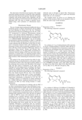
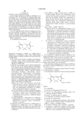
- the acidic organelle stains of the present invention are dipyrrometheneboron difluoride dyes having the formula ##STR1## where the substituents R 1 -R 6 , which may be the same or different, are hydrogen, halogen, cyano, alkyl, perfluoroalkyl, alkoxy, cycloalkyl, arylalkyl, acyl, aryl, heteroaryl, alkenyl or alkynyl; or a LINK-CAP moiety.
- the bridging R 7 is optionally a nitrogen atom, yielding an aza-dipyrrometheneboron difluoride dye, or R 7 is a methine (--CH ⁇ ) or a methine that is substituted by halomethyl, cyano, alkyl, perfluoroalky, alkenyl, alkynyl, cycloalkyl, arylalkyl, acyl, aryl, heteroaryl, or a LINK-CAP moiety.
- R 7 is a methine or substituted methine.
- R 7 is a substituted methine, preferably the substituents are halomethyl or a LINK-CAP moiety.
- the substituents R 1 -R 6 that are not a LINK-CAP moiety are hydrogen, halogen, alkyl, aryl, heteroaryl or alkenyl. More preferably, the substituents R 1 -R 6 that are not a LINK-CAP are hydrogen, halogen or alkyl.
- any two adjacent substituents of R 1 , R 2 , R 3 , R 4 , R 5 and R 6 taken in combination, form a fused aromatic 6-membered ring that is optionally and independently substituted one or more time, at any position, by halogen, cyano, alkyl, perfluoroalkyl, alkoxy, alkenyl, alkynyl, cycloalkyl, alkylthio, alkylamido, amino, monoalkylamino, dialkylamino, carboxamide, hydroxy, mercapto, aryl, heteroaryl, aryl-amido, heteroaryl-amido, aryl-oxy, heteroaryl-oxy, aryl-amino, or heteroaryl-amino, or 1-2 additional fused benzo or heteroaromatic rings that are themselves optionally further substituted by halogen, amino or carboxamide. Any of the fused aromatic 6-membered
- aryl is an aromatic or polyaromatic substituent containing 1 to 4 aromatic rings (each ring containing 6 conjugated carbon atoms and no heteroatoms) that are optionally fused to each other or bonded to each other by carbon-carbon single bonds. Each aryl is bound to the dye by a single bond, and is optionally substituted as described below.
- a heteroaryl is an aromatic group that contains at least one heteroatom (a non-carbon atom within the ring structure).
- Each heteroaryl is a single 5- or 6-member ring, or is a fused 2- or 3-ring structure.
- the heteroaryl group contains one or more heteroatoms, e.g. as pyrrole, thiophene, or furan (single ring, single heteroatom), or oxazole, isoxazole, oxadiazole, or imidazole (single ring, multiple heteroatoms), or benzoxazole, benzothiazole, or benzimidazole (multi-ring, multiple heteroatoms), or benzofuran or indole (multi-ring, single heteroatom).
- Each heteroaryl is bound to the dye by a single bond, and is optionally substituted as described below.
- alkenyl or alkynyl substituents independently has 2-6 carbons, and is optionally substituted by halogen, alkyl, cyano, carboxylate ester, carboxamide, aryl, heteroaryl, or additional alkenyl or alkynyl groups.
- an alkenyl group is an ethenyl, dienyl or trienyl group.
- Each of the alkyl substituents, as well as the alkyl portions of alkoxy, cycloalkyl, arylalkyl, alkylamino, alkylthio or alkylamido substituents independently has 1-6 carbons, and is optionally substituted by halogen, amino, alkylamino, dialkylamino, carboxamide, hydroxy, mercapto or cyano.
- At least one of R 1 -R 6 is a LINK-CAP moiety, or R 7 is a LINK-CAP substituted methine, or one of the dye substituents that is a fused 6-membered ring is further substituted by a LINK-CAP moiety.
- the dye is substituted by more than one LINK-CAP, they are the same or different.
- the LINK portion of LINK-CAP is a covalent linkage, serving to attach a weakly basic amine, CAP, to the dipyrrometheneboron difluoride fluorophore itself. Any suitable covalent linkage that does not interfere with the ability of the dye to selectively accumulate in acidic organelles is an acceptable covalent linkage for the purposes of the present invention.
- LINK is a single covalent bond.
- Preferred LINK groups have 1-20 nonhydrogen atoms selected from the group consisting of C, N, O and S.
- Such LINK groups are composed of any combination of chemical bonds, including ether, thioether, amine, ester, carboxamide, sulfonamide, hydrazide bonds, and single, double, triple carbon-carbon bonds, and aromatic or heteroaromatic bonds.
- Preferred LINK groups are composed of any combination of single carbon-carbon bonds and carboxamide bonds.
- Selected specific examples of LINK optionally include methylenes, oligomethylenes, phenylenes, thienyls, carboxamides, and sulfonamides.
- LINK contains 1-6 carbon atoms.
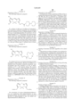
- LINK has the formula --(CH 2 ) a (CONH(CH 2 ) b ) z --, where a has any value from 0-5, b has any value from 1-5 and z is 0 or 1.
- CAP has the general formula --CR 8 R 9 --NR 10 R 11 .
- the substituents R 8 and R 9 are independently hydrogen or alkyls having 1-6 carbons that are linear or branched. Typically, R 8 and R 9 are hydrogen or alkyls having 1-2 carbons, more preferably both R 8 and R 9 are hydrogen. Where R 8 or R 9 are alkyl groups, each alkyl group is optionally further substituted by halogen, carboxamide, hydroxy, mercapto or cyano. In addition, any alkyl group is optionally further substituted by a primary, secondary or tertiary amine, where the alkyl groups present on the amine independently have 1-6 carbons. In an additional embodiment of the invention, one of R 8 and R 9 , when taken in combination with the LINK moiety, forms a six- to eight-membered ring.
- the amine substituents R 10 and R 11 are each independently H or a linear or branched alkyl having 1-6 carbons.
- R 10 and R 11 when taken in combination form a nitrogen heterocycle that preserves the basic nature of the amine nitrogen.
- the nitrogen heterocycle is a pyrrolidine, a piperidine, a piperazine, morpholine, an imidazole, an azepine (including diazepines and triazepines) or an oxazepine.
- the amine substituents R 10 and R 11 when taken in combination with substituents R 8 and R 9 , or with the LINK moiety, form a saturated five- or six-membered nitrogen heterocycle that is a substituted pyrrolidine or piperidine.
- the basic CAP moiety is optionally present in the form of a salt of a strong acid, for example the hydrochloride salt, sulfate salt, perchlorate salt, or other organic acid salts.
- the dyes of the present invention are readily prepared using the methods described in U.S. Pat. No. 4,774,339 to Haugland, et al. (1988), U.S. Pat. No. 5,187,288 to Kang et al. (1993), U.S. Pat. No. 5,248,782 to Haugland, et al. (1993), U.S. Pat. No. 5,274,113 to Kang, et al. (1993), and U.S. Pat. No. 5,433,896 to Kang, et al. (1995) (all incorporated by reference).
- Methods for preparing aza-substituted dipyrrometheneboron difluoride dyes are described in U.S. Pat. No.
- Compounds wherein the LINK or CAP moiety incorporates a cyclic structure are prepared by reaction of a preformed reactive dye with an appropriate amine-containing intermediate, or by preparing amine-containing pyrroles prior to formation of the dye.
- the dyes of the invention are only partially protonated at neutral pH.
- the spectral properties of the probe can be tuned over a wide range of the visible and near infra-red spectrum, making them especially useful for multicolor applications.
- careful selection of substituents allow the pH selectivity of the dyes of the invention to be tuned for specific applications. For example, a dye having a CAP moiety that is less basic will be protonated by more acidic conditions, and therefore more selectively accumulate at locations having a lower pH.
- the preferred dyes of the present invention are freely permeant to cell membranes, and typically selectively accumulate in acidic organelles.
- the staining characteristics are generally not reversed or are only partially reversed by subsequent treatment of the cells with additional weakly basic cell-permeant compounds. In some cases, the staining is preserved even after fixation and/or permeabilization of the cells.
- the probes of the present invention are utilized by preparing a labeling solution containing one or more of the dyes of the present application, introducing the labeling solution into the sample containing or thought to contain acidic organelles, incubating the sample for a time sufficient to produce a detectable fluorescent staining pattern, and observing or analyzing the staining pattern in the sample.
- the sample may be a cell or cells that contain acidic organelles or the sample may contain isolated acidic organelles (i.e. not incorporated in a cell), or the sample may be two solutions separated by a semi-permeable membrane.
- the degree of staining of acidic organelles is a reflection of the pH gradient present across the acidic organelles membrane at the time of staining, i.e., the degree of staining is indicative of whether or not the organelle is acidic at the time of staining.
- the dyes of the present invention are typically used for staining the acidic organelles of live cells, the present invention is also useful for staining isolated (i.e. cell-free) acidic organelles, provided the organelles are not disrupted and a pH gradient still exists between the organelle and the medium in which it is suspended. While in general the presence of acidic organelles can be considered an indicator of cell viability, it is possible to render a cell non-viable, while still retaining acidic organelles in the sample.
- the pure dyes generally have low solubility in water.
- a stock solution is prepared by dissolving a known mass of the pure dye in an organic solvent.
- organic solvents are DMSO, DMF, N-methylpyrrolidone, acetone, acetonitrile, dioxane, tetrahydrofuran and other nonhydroxylic, completely water-miscible solvents.
- the dye is dispersed in a water immiscible solvent or oil, or is evaporated from an organic solvent leaving a thin film.
- the stock solutions should be protected from light at all times.
- the labeling solution is prepared by diluting an aliquot of the stock solution into an aqueous or partially aqueous buffer to the desired labeling concentration.
- two or more dyes of the invention are present in the labeling solution, having similar or distinct spectral properties.
- the amount of dye added in the labeling solution is the minimum amount required to yield detectable staining of the acidic organelles present in the sample within a reasonable time, with minimal background fluorescence or staining of other organelles or cellular structures.
- the amount of dye required for staining eukaryotic cells depends on the sensitivity required for staining the intracellular acidic organelles, the number of cells present, the permeability of the cell membrane to the dye, and the time required for the probe to localize to the organelles.
- the required concentration for the labeling solution is determined by systematic variation in labeling concentration until a satisfactory fluorescent labeling is accomplished.
- the amount of dye required for staining animal cells is 20 to 400 nM, preferably below 100 nM.
- Staining concentrations less than about 100 nM give good staining of acidic organelles in live animal cells. At higher concentrations of stain, background fluorescence increase in live cells, but resolution of acidic organelles after fixation is improved. Staining of isolated (cell-free) acidic organelles typically requires lower concentrations of dye.
- concentration of stain to be used is dependent upon the experimental conditions and the desired results and optimization of experimental conditions is required to determine the best concentration of stain to be used in a given application.
- the sample optionally comprises cell-free acidic organelles or cells that contain acidic organelles.
- Any cells that contain acidic organelles can be used, including but not limited to, fresh or cultured cells, cell lines, cells in biological fluids, cells in tissue or biopsy, yeast cells, plant cells and sperm cells.
- the cells are optionally abnormal cells, such as tumor cells or other cancer cells, where the abnormal cells are present in vitro or in vivo.
- Acidic organelles of interest that are stained using the present method of staining include, but are not limited to, lysosomes, phagovacuoles, endosomes, yeast vacuoles and acrosomes.
- the staining method is used to stain all lysosomal compartments in the sample.
- the acidic organelles that are stained are lysosomes or acrosomes. More typically, the acidic organelles that are stained are lysosomes.
- vacuoles Most plant and fungal cells (including the unicellular fungi and yeast) contain one or more very large, fluid-filled vesicles called vacuoles. In yeast, the vacuoles typically occupy more than 70% of the cell volume. Yeast vacuoles are related to lysosomes of animal cells, and contain a variety of hydrolytic enzymes with acidic pH in the lumen.
- Mature sperm cells are relatively simple cells, containing only a cell membrane, a compact nucleus and a vesicle, called an acrosome.
- the acrosome is derived from a Golgi body and is located at the leading tip of the sperm cell.
- the acrosome is an acidic compartment containing enzymes that aid in the penetration of the protective layers surrounding egg, initiating the egg activation step (the first step in the fertilization process). Therefore, acrosomes play a very important role in egg fertilization.
- the method of the present invention is useful for the staining and monitoring of sperm acrosomes. Only healthy sperm cells with acidic acrosomes are stained. The resulting stained sperm cells can then be sorted using flow cytometry or another fluorescence-sensitive device in order to improve fertilization rate, or to conduct research.
- the sample is typically stained by passive means, that is the labeling solution is combined with the sample being analyzed.
- the dyes of the present invention are introduced into the sample organelles by incubation of the sample in the labeling solution. Where the sample contains a cell or cells, the cells are similarly stained by incubation of the cell or cells in the labeling solution. Alternatively, the sample is stained by direct uptake of the dye from a thin film of the dye itself. Any other method of introducing the dye into the sample cell, such as microinjection of a labeling solution, can be used to accelerate introduction of the dye into the cellular cytoplasm. Typically the dye will be introduced into the sample cell by incubation in the labeling solution, or by microinjection. Preferably the dye is introduced to the sample by incubation in the labeling solution. Microinjection of dye solution is used when labeling of the acidic organelles in a single cell is desired, within a colony of other sample cells.
- a number of reagents and conditions are known to affect the pH gradient of acidic organelles, and therefore the staining of the dyes of the invention, including but not limited to nutrients (for example carbohydrates such as glucose) and selected drugs.
- the dyes of the present invention are generally non-toxic to living cells and acidic organelles. Sample cells have been incubated in 75 nM dye solution for 36 hours without observable ill effects. Stained cells have been observed to undergo cell division, producing daughter cells that also possess stained acidic organelles.
- the cells or isolated acidic organelles are washed to improve the results of the staining procedure. Washing the sample cell or cells after incubation in the labeling solution, or optionally after fixation or permeabilization, greatly improves the visualization of the acidic organelles. This is largely due to the decrease in non-specific background fluorescence after washing. Satisfactory visualization of acidic organelles is possible without washing by using low labeling concentrations (for example ⁇ 50 nM).
- the sample can be observed immediately after staining of acidic organelles is evident.
- the cells or isolated acidic organelles in a sample are optionally fixed.
- Selected embodiments of the dyes described above are well retained in cells, and sample cells stained with these dyes retain considerable fluorescent staining after fixation.
- fixatives and fixation conditions are suitable for practicing this invention.
- Useful fixatives include, but are not limited to, formaldehyde, paraformaldehyde, formalin, glutaraldehyde, cold methanol and 3:1 methanol acetic acid.
- cell fixation is accomplished by incubating the stained cells in a 3.7% solution of paraformaldehyde for about 15-30 minutes. Fixation is typically used to preserve cellular morphology and to reduce biohazards when working with pathogenic samples.
- Fixation is optionally followed or accompanied by permeabilization, such as with acetone, ethanol, DMSO or various detergents. Permeabilization is utilized to allow bulky additional detection reagents to enter the cellular space that would ordinarily be impermeant to an intact cellular membrane.
- permeabilization such as with acetone, ethanol, DMSO or various detergents.
- Permeabilization is utilized to allow bulky additional detection reagents to enter the cellular space that would ordinarily be impermeant to an intact cellular membrane.
- fixatives, fixation conditions, and permeabilization agents are known in the art, and other methods of fixing or permeabilizing sample cells in conjunction with the stains of the present invention will be obvious to one of ordinary skill.
- An additional detection reagent is a reagent that produces a detectable response due to the presence of a specific cell component, intracellular substance, or cellular condition.
- One or more additional detection reagents may be used in conjunction with the stains of the present invention, before or after fixation and/or permeabilization.
- the additional detection reagent may be used to stain the entire cell, or a cellular substructure.
- the fluorescent signal of the acidic organelle stains of the present invention and the detectable response of the additional detection reagent may be observed simultaneously or sequentially.
- nucleic acid stains One class of appropriate additional detection reagents are fluorescent nucleic acid stains.
- a wide variety of appropriate nucleic acid stains are known in the art, including but not limited to, Thiazole Orange, ethidium homodimer, propidium iodide, Hoechst 33258 (Example 21), and DAPI. Additional useful nucleic acid stains are described in the international applications WO 93/06482, DIMERS OF UNSYMMETRICAL CYANINE DYES (published Apr. 1, 1993); U.S. Pat. No. 5,436,134 to Haugland et al., 1995; U.S. Pat. No. 5,321,130 to Yue et al, 1994; U.S. Pat. No.
- nucleic acid stain in conjunction with the dyes of the present invention can be selected to allow simultaneous observation of acidic organelles, nuclear DNA, cellular RNA and/or mitochondrial DNA.
- an additional detection reagent that is a cell-permeant nucleic acid stain, such as those described in U.S. Pat. No. 5,436,134, allowing simultaneous visualization of acidic organelles and the cell nucleus.
- additional detection reagents include selected fluorescent metal ion indicators described in U.S. Pat. No. 5,453,517 to Kuhn et al. (1995), or U.S. Pat. No. 5,405,975 to Kuhn et al. (1995).
- an appropriate additional detection reagent is any probe that selectively stains a cellular organelle such as the cell membrane, nucleus, Golgi apparatus, mitochondrion, endoplasmic reticulum, or is a second acidic organelle probe.
- additional detection reagents include mitochondria-targeted stains, such as Rhodamine 123.
- Additional fluorescent stains specific for mitochondria are described in U.S. Pat. No. 5,459,268 to Haugland et al. (1995) (hereby incorporated by reference).
- the above mitochondrial stains accumulate in mitochondria, and are fixable therein, allowing simultaneous visualization of both mitochondria and acidic organelles in fixed and permeabilized cells.
- the additional detection reagent comprises: a) one member of a specific binding pair or a series of specific binding pairs, and b) a means for producing a detectable response.
- a specific binding pair member can be a ligand or a receptor.
- ligand means any organic compound for which a receptor naturally exists or can be prepared.
- a receptor is any compound or composition capable or recognizing a spatial or polar organization of a molecule, e.g. epitopic or determinant site.
- Ligands for which naturally occurring receptors exist include natural and synthetic peptides and proteins, including avidin and streptavidin, antibodies, enzymes, and hormones; nucleotides and natural or synthetic oligonucleotides; lipids; polysaccharides and carbohydrates; lectins; and a variety of drugs, including therapeutic drugs and drugs of abuse and pesticides.
- Ligands and receptors are complementary members of a specific binding pair, each specific binding pair member having an area on the surface or in a cavity which specifically binds to and is complementary with a particular spatial and polar organization of the other.
- the additional detection reagent may be used in conjunction with enzyme conjugates to localize cellular receptors; to localize hybridization probes; or to probe cells and tissues that do not express the enzyme, for example, by enzyme-linked immunosorbent assay (ELISA), or enzyme-mediated histochemistry or cytochemistry, or other enzyme-mediated techniques. Enzyme-mediated techniques take advantage of the attraction between specific binding pairs to detect a variety of analytes.
- the additional detection reaction comprises an enzyme substrate to produces a fluorescent precipitate in the presence of the appropriate enzyme, as described in U.S. Pat. No. 5,316,906 to Haugland et al. (1994) and U.S. Pat. No. 5,443,986 to Haugland et al. (1995).
- an enzyme-mediated technique uses an enzyme attached to one member of a specific binding pair or series of specific binding pairs as a reagent to detect the complementary member of the pair or series of pairs.
- One member of the specific binding pair is the analyte, i.e. the substance of analytical interest.
- An enzyme is attached to the other (complementary) member of the pair, forming a "complementary conjugate".
- multiple specific binding pairs may be sequentially linked to the analyte, the complementary conjugate, or to both, resulting in a series of specific binding pairs interposed between the analyte and the detectable enzyme of the complementary conjugate incorporated in the specific binding complex.
- the additional detection reagent also incorporates a means for producing a detectable response.
- a detectable response means a change in, or occurrence of, a parameter in a test system which is capable of being perceived, either by direct observation or instrumentally, and which is a function of the presence of a specifically targeted member of a specific binding pair in a cell sample.
- detectable responses include the change in, or appearance of, color, fluorescence, reflectance, pH, chemiluminescence, infrared emission, or the deposition of an electron-rich substrate.
- Appropriate labels to provide a detectable response include, but are not limited to, a visible or fluorescent dye, an enzyme substrate which produces a visible or fluorescent precipitate upon enzyme action (for example, the action of horseradish peroxidase upon diaminobenzidine), visible or fluorescent labeled latex microparticles, or a signal that is released by the action of light upon the reagent (e.g. a caged fluorophore that is activated by photolysis, or the action of light upon diaminobenzidine).
- a visible or fluorescent dye for example, the action of horseradish peroxidase upon diaminobenzidine
- an enzyme substrate which produces a visible or fluorescent precipitate upon enzyme action
- visible or fluorescent labeled latex microparticles for example, the action of horseradish peroxidase upon diaminobenzidine
- a signal that is released by the action of light upon the reagent e.g. a caged fluorophore that is activated by photolysis, or the action
- the sample is illuminated with a wavelength of light that results in a detectable fluorescence response, and subsequently observed with a means for detecting the detectable response of fluorescent labeled acidic organelles, if present.
- the fluorescently labeled organelles are observed after the cell or cells have additionally been fixed and/or permeabilized. Observation is accomplished using visible light microscopy, or alternatively, observation of the sample comprises illuminating the stained sample with a wavelength of light appropriate to generate a fluorescent response, and visually examining the sample by use of a microscope, or confocal microscope.
- the sample is optionally illuminated at a wavelength specific for optimal excitation of a single dye present in the sample.
- illumination occurs at a wavelength that generates a detectable fluorescence response in each dye or additional detection reagent, where said dyes and detection reagents possess overlapping excitation maxima.
- the dyes of the invention typically possess a strong absorbance at visible wavelengths, typically at greater than 450 nm, preferably at greater than 600 nm, yet more preferably at greater than 650 nm.
- the preferred dyes of the invention exhibit an extinction coefficient greater than 50,000 cm -1 M -1 , preferably at greater than 80,000 cm -1 M -1 .
- the dyes of the invention typically possess quantum yields of fluorescence emission that are greater than 0.3, preferably greater than 0.7.
- the sample is observed using instrumentation.
- observation of the sample is accomplished by illuminating the stained cell or cells with a wavelength of light appropriate to generate a fluorescent response, and electronically detecting and optionally quantifying the fluorescent emission of the stained acidic organelles using an appropriate instrument, such as a fluorometer, fluorescent microplate reader, or a flow cytometer.
- the observation of the fluorescent response of the sample optionally includes selecting or sorting the acidic organelles based upon their fluorescent response.
- the sample comprises cells having stained acidic organelles, and the cells of the sample are sorted based upon the staining of the individual cells.
- the step of sorting is typically accomplished using a flow cytometer or a fluorescence microscope.
- Photodynamic therapy refers to the process wherein illumination is utilized to destroy cells, typically abnormal cells, that have previously been labeled with a dye.
- CANCER 59, 814-822 (1994); incorporated by reference) have previously indicated that the photolysis of dyes that are localized to lysosomes destroys tumor cells. Furthermore, the lysosomes of tumor cells are generally considered to have a lower pH than normal lysosomes ("Molecular Aspects of Anticancer Drug Action", Neidel and Waring, Eds., Macmillan, London; pp 233-286 (1983), incorporated by reference). Selective uptake of PDT dyes into tumor cells in preference to normal cells is an important property allows selective photodestruction of abnormal cells in the course of PDT treatment, while minimizing the destruction of normal cells.
- the method of the current invention has utility for photodynamic therapy, as described above, as the greater acidity of lysosomes in tumor cells, will result in greater uptake of the acidotropic dyes in tumor cells. Photolysis of the stained cells will then result in destruction of the target cells.
- cells and tissues stained according to the present method are potential PDT targets, preferably the dipyrrometheneborondifluoride dyes used for PDT targeting of cells are those that absorb beyond 600 nm, more preferably those that absorb beyond 650 nm, due to the enhanced penetration of light through tissue at these wavelengths.
- Particularly preferred are the dyes of the invention having fused benzo substituents, as described in U.S. Pat. No. 5,433,896 to Kang et al. (1995) that are further substituted by a LINK-CAP moiety.
- Additional preferred dyes of the invention for PDT are those having bromine or iodine substituents.
- Preferred dye concentrations for labeling cells for PDT are those concentrations that have been determined to produce the greatest selective uptake of dye into abnormal cells without detriment to normal cells, such that photolytic activity is maintained in the abnormal cells.
- submicromolar concentrations of dye are effective in staining acidic organelles of live cells.
- the dyes are applied to cells for PDT by means well understood in the art, including local or systemic injection, topical application, incorporation into liposomes or other means.
- Dye uptake into cells is by passive diffusion or receptor-mediated uptake. Selective accumulation in lysosomes is facilitated by the pH gradient that favors uptake into the more acidic organelles.
- Photolysis is performed with any excitation source that is capable of producing light that can be absorbed by the dye, including lasers and light sources that produce infrared irradiation.
- This light may be delivered either directly to the cells that contain the dye, or delivered indirectly such as through an optical fiber. Fluorescence properties of the dye can be used to guide and determine which cells are to be irradiated.
- succinimidyl ester (0.25 mmoles) in 10 mL of dichloromethane is dropwise added 22.0 mg of N,N-dimethylethylenediamine (0.25 mmoles) in 10 mL of dichloromethane at 0° C. under stirring. The mixture is warmed up to room temperature, and continuously stirred for 6 hours. The resulted solution is diluted using chloroform, and washed with water.
- succinimidyl ester (0.23 mmoles) in 10 mL of dichloromethane is dropwise added 22.0 mg of N,N-dimethylethylenediamine (0.25 mmoles) in 10 mL of dichloromethane at 0° C. under stirring. The mixture is warmed to room temperature, and continuously stirred for 6 hours. The resulted solution is diluted using chloroform, and washed with water.
- the chloroform layer is dried over anhydrous sodium sulfate, and evaporated in vacuo to afford red solid.
- the crude product of 2,6-dibromo-4,4-difluoro-5,7-dimethyl-4-bora-3a,4a-diaza-s-indacene-3-propionic acid is obtained.
- the crude product is purified by silica gel column chromatography using 3% methanol in chloroform as eluant to give 57 mg of the pure product.
- a sample of 20 mg of the acid is converted to a reactive succinimidyl ester as follows: To a solution of 20 mg of the above acid in 10 mL of ethyl acetate is added 7 mg of N-hydroxysuccinimide, followed by addition of 10 mg of N,N-dicyclohexylcarbodiimide and the mixture is stirred at room temperature for 3 hours. The reaction mixture is concentrated under reduced pressure and is subjected to silica gel column using chloroform as eluant to give 20 mg of the reactive succinimidyl ester derivative.
- succinimidyl ester (20 mg) is added to a solution of 30 ⁇ L of N,N-dimethylethylenediamine in 5 mL of dichloromethane. After the mixture is stirred at room temperature for 1 hour, it was subjected to silica gel column chromatography using 10% methanol in chloroform. The desired fractions from the column are combined and treated with 200 ⁇ L of 4M HCl solution. The resulting solution is concentrated under reduced pressure and the residue is applied to a short SEPHADEX LH-20 resin column and eluted with water. The desired fractions arc combined and lyophilized to give the desired product as the hydrochloride salt.
- the desired fractions from the column are combined and treated with 300 ⁇ L of 4M HCl solution.
- the resulting solution is concentrated under reduced pressure and the resulting residue is applied to a SEPHADEX LH-20 resin column and eluted with water. After lyophilization of the desired combined fractions, the desired product is obtained.
- reaction mixture After stirring at room temperature for 1 hour, the reaction mixture is purified by silica gel column chromatography using 15% methanol in chloroform as eluant. The desired fractions are combined, and treated with 300 ⁇ L of 4M HCl solution. The resulting solution is concentrated under reduced pressure and the resulting residue is subjected to SEPHADEX LH-20 resin column and eluted with water. After lyophilization, the desired product is obtained.
- Dye 54 is prepared analogously to Dye 21 above (Example 3), excepting that 4,4-difluoro-1,3,5,7-tetramethyl-2-iodoacetamido-4-bora-3a,4a-diaza-s-indacene is used in place of 4,4-difluoro-5,7-dimethyl-3-iodoacetamidomethyl-4-bora-3a,4a-diaza-s-indacene.
- a calf pulmonary arterial endothelium (CPAE) cell line is obtained from American Type Culture Collection Co., Rockville Md.
- the cells are maintained in a humidified atmosphere of 5% CO 2 in Dulbecco's modified Eagle's medium (DMEM) supplemented with 10% calf serum, 50 ⁇ g/mL gentamicin, 300 ⁇ g/mL L-glutamime and 10 mM HEPES pH 7.4.
- Cells are subcultured every 2-3 days by trypsinization using 0.05% trypsin and 0.02% EDTA in a calcium- and magnesium-free saline solution (Gibco BRL, Gaithersburg, Md.).
- DMEM Dulbecco's modified Eagle's medium
- EDTA calcium- and magnesium-free saline solution
- the desired dye of the invention is separately dissolved in DMSO to prepare a 1 mM dye stock solution.
- the stock solution is kept sealed in small aliquots, at -20° C.
- the stock solution is kept frozen at all times until use, and exposure to light is minimized.
- One aliquot of dye stock is taken from the freezer immediately before an experiment and thawed completely at room temperature.
- the labeling solution is then prepared by adding the dye stock solution to fresh culture medium in an amount sufficient to make final dye concentrations of 50 and 200 nM, respectively.
- Example 19 Cells prepared according to Example 19 are transferred to the labeling solution containing Dye 40 (Example 8), prepared according to Example 20, and incubated at 37° C. for 20 to 60 minutes. The cells are then washed with pre-warmed fresh medium that does not contain phenol red (37° C.) and observed using a Zeiss Axioplan epifluorescence microscope equipped with an appropriate filter set.
- Cells prepared according to Example 19 are transfered to the labeling solution containing 75 nM Dye 3 and 75 nM of the mitochondrial stain MitoTracker CMXRos (Molecular Probes, Eugene Oreg.) and incubated at 37° C. for 30 minutes. The cells are then washed with fresh, pre-warmed culture medium that does not contain phenol red and examined under an epifluorescence microscope equipped with multibandpass filter set, such as an XF56 filter set (Omega Optical). As both dyes are organelle-specific, the lysosomes and other acidic organelles stain are stained a bright fluorescent green, while the mitochondria are simultaneously stained fluorescent red. No cross staining of organelles, indicated by a yellow fluorescent signal, is observed.
- CMXRos Molecular Probes, Eugene Oreg.
- a sample of fungal cells is resuspended at 10 6 cells/mL in 10 mL HEPES buffer, pH7.4, containing 5% glucose.
- a labeling solution of the desired dye (as prepared in Example 19) is added to the fungal cell suspension, to a final dye concentration of 10 ⁇ M.
- the cells are incubated at room temperature for a maximum of 3 to 5 minutes.
- the stained cells are pelleted by centrifugation and resuspended in fresh 10 mM HEPES buffer, pH 7.4, containing 5% glucose.
- the cells are then examined with a Zeiss microscope equiped with an appropriate filter set.
- the commonly used vacuole marker, 2',7'-dichlorodihydrofluorescein diacetate can be used as a control for verifying the specificity of vacuolar staining.
- a semen sample is diluted in HEPES-buffered isotonic saline solution containing serum albumin (10 mM HEPES, 0.85% NaCl, 10% BSA, pH 7.4). The sample is kept at 36° C. A 1 ⁇ M stock solution of the desired dye is prepared in the same buffer. A 100 ⁇ L aliquot of the stock solution is added to 900 ⁇ L of diluted semen, resulting in a final dye concentration of 100 nM. The sample is incubated with the dye for 20-30 minutes at 36° C. The sample is then observed using a fluorescent microscope equipped with a proper filter set (based on the excitation/emission wavelength of the dye used). Alternatively, the sample is analyzed and sorted using flow cytometry.
- CPAE cells are incubated for 30 minutes in a solution of culture medium and 100 ⁇ g/mL Datura stramonium (jimson weed) lectin conjugated to fluorescein at 37° C. in a tissue culture incubator. This green fluorescent lectin has been shown to be taken up by live cells and trafficked to lysosomes.
- the cells are rinsed twice in fresh culture medium at 37° C., and then are allowed to recover in culture medium without lectin for 4 hours at 37° C. Following recovery, the cells are incubated with 100 nM Dye 43 and diluted in growth medium for 30 minutes at 37° C. The cells are then rinsed in growth medium without phenol red and mounted in the same.
- the observed yellow fluorescence indicates colocalization of the green fluorescent lectin conjugate with the red dye of the invention in lysosomes.
- the red structures indicate the staining of acidic organelles that are not lysosomes.
- lysosomes are the principal sites of intracellular digestion, there are some 40 acidic hydrolytic enzymes contained in lysosomes. There are also a plurality of different proteins present on the lysosomal membrane that play important roles in critical processes, including material transportation and H + pumping into the lumen. Both classes of lysosomal proteins are synthesized in the rough endoplasmic reticulum and transported through the Golgi apparatus. The precursors of all lysosomal hydrolases therefore carry a unique marker in the form of a mannose 6-phosphate (M6P) group, which will target the proteins for transport to lysosomes specifically.
- M6P mannose 6-phosphate
- a researcher can clone a gene sequence and manipulate it in such a way that the final product, a polypeptide, will fuse with the N-terminal first 100 amino acid residues of a regular lysosomal hydrolytic enzyme (L1).
- This fusion protein should have a M6P group and therefore target the lysosomes in transformed cells in a culture.
- the fusion protein can then be detected using immunocytochemical methods in combination with the dyes of the present invention.
- the typical procedure for the expression and detection of a foreign gene products in lysosomes of transformed cells comprises the following steps:
- a specific protein with a known amino acid sequence for example, a subunit of yeast protein: T1 is selected, and monoclonal antibodies (McAb) directed against that sequence are prepared.
- the selected DNA sequence coded for Y1 from a yeast cDNA library is amplified using the polymerase-chain reaction.
- the selected DNA sequence is then fused with amplified, selected bovine nuclear DNA, which codes for the first 100 amino acids of a bovine lysosomal protein (L1).
- L1 bovine lysosomal protein
- the fused gene is ligated into a mammalian expression vector, such as pCI (Promega), which has the proper enhancer/promoter and other signal regions for accurate, high efficiency gene expression in bovine cells.
- a mammalian transfection system is used to transfer the gene construct into cultured bovine cells, establishing a stable, transformed cell line that expresses lysosomal targeting polypeptide T1/L1. The transformed cells are cultured under proper conditions.
- the cells are stained with 150 nM of Dye 43 at 37° C. for 30 minutes.
- the lysosomes in the living cells are well labeled and fluoresce bright red.
- the stained cells are then fixed in 3.0% formaldehyde/0.5% glutaraldehyde for 20 min at room temperature and permeabilized with cold acetone for 10 min.
- Dye 43 is well retained in lysosomes throughout this cytochemical process.
- the sample cells are further incubated with McAb against T1.
- the immunocomplexes are detected with FITC-labeled secondary antibodies. Those regions of the sample showing both red fluorescence (lysosomes) and green fluorescence (immunocomplexes) indicate the antibody complex colocalizes in the lysosomes of the sample.
- Cell samples prepared according to Example 19 are transferred to two tissue culture dishes.
- the cells in the first dish are pre-incubated with 1 mM NH 4 OH for 30 minutes at 37° C.
- the cells are then washed with fresh medium.
- the cells in both dishes are then incubated with a labeling solution that is 75 nM in Dye 3 (as described in Example 2) at 37° C. for 30 minutes.
- the cells are then washed with pre-warmed fresh medium and observed using a Zeiss Axioplan microscope equipped with an appropriate filter set.
- the cells that were not initially alkalinized display good staining, with all of the lysosomes and other acidic organelles exhibiting a bright green fluorescence.
- the initially alkalinized cells show only weak staining of acidic organelles, displaying less than 10% of the fluorescence intensity of the control cells.
- a cell sample prepared according to Example 19 are transferred to a tissue culture dish and incubated at 37° C. for 30 minutes in a labeling solution that is 75 nM in Dye 3 (as described in Example 2), 100 nM in BODIPY TR labeled ceramide (Molecular Probes, Inc., Eugene Oreg.) and 30 nM in Hoechst 33258 (Molecular Probes, Inc., Eugene Oreg.).
- the cells are then washed with fresh, pre-warned culture medium and examined under a Zeiss Axioplan microscope equipped with an appropriate filter set.
- the stained cells display green fluorescent acidic organelles, red fluorescent Golgi apparatus, and blue fluorescent nuclei.
- a cell sample prepared according to Example 19 is incubated at 37° C. for 30 minutes in a labeling solution that is 75 nM in Dye 3 and 50 nM in propidium iodide.
- the stained cells are then washed with fresh, pre-warmed culture medium.
- the cells are examined under a Zeiss Axioplan microscope equipped with an appropriate filter set. Dead cells exhibit fluorescent red nuclei, while live cells exhibit green fluorescent acidic organelles. Cells that have damaged (i.e. permeant) cell membranes, yet retain an acidic pH gradient with their acidic organelles will display both green and red fluorescence.
Abstract
Description
TABLE 1
__________________________________________________________________________
Selected Embodiments of the Dyes of the Invention
##STR2##
Dye
No.
R.sup.1 R.sup.2
R.sup.3 R.sup.4 R.sup.5
R.sup.6
R.sup.7
__________________________________________________________________________
1 H H (CH.sub.2).sub.2 CONH
Me H Me CH
(CH.sub.2).sub.2 NH.sub.2
2 H H (CH.sub.2).sub.2 CONH
Me H Me CH
(CH.sub.2).sub.2 NHMe
3 H H (CH.sub.2).sub.2 CONH
Me H Me CH
(CH.sub.2).sub.2 NMe.sub.2
4 H H (CH.sub.2).sub.2 CONH
Me H Me CH
(CH.sub.2).sub.2 N(Et).sub.2
5 H H (CH.sub.2).sub.2 CONH
Me H Me CH
(CH.sub.2).sub.2 N(Pr).sub.2
6 H H (CH.sub.2).sub.2 CONH
Me H Me CH
(CH.sub.2).sub.2 N(Bu).sub.2
7 H H
##STR3## Me H Me CH
8 H H (CH.sub.2).sub.2 SO.sub.2 NH
Me H Me CH
(CH.sub.2).sub.2 NH.sub.2
9 H H (CH.sub.2).sub.2 SO.sub.2 NH
Me H Me CH
(CH.sub.2).sub.2 N(Me).sub.2
10 H H (CH.sub.2).sub.3 NH.sub.2
Me H Me CH
11 H H CH.sub.2 N(Me).sub.2
Me H Me CH
12 H H CH.sub.2 NH.sub.2
Me H Me CH
13 H H (CH.sub.2).sub.3 NH
Me H Me CH
(CH.sub.2).sub.3 N(Me).sub.2
14 H H (CH.sub.2).sub.3 NH
Me H Me CH
(CH.sub.2).sub.2 N(Bu).sub.2
15 H H (CH.sub.2).sub.3 NHCSNH
Me H Me CH
(CH.sub.2).sub.2 N(Me).sub.2
16 H H (CH.sub.2).sub.2 COO
Me H Me CH
(CH.sub.2).sub.2 N(Me).sub.2
17 H H (CH.sub.2).sub.3 NHCONH
Me H Me CH
(CH.sub.2).sub.2 N(Me).sub.2
18 H H (CH.sub.2).sub.2 CONH(CH.sub.2).sub.5
Me H Me CH
CONH(CH.sub.2).sub.2 N(Me).sub.2
19 H H (CH.sub.2).sub.2 CONH(CH.sub.2).sub.2 NH
Me H Me CH
COCH.sub.2 S(CH.sub.2).sub.2 N(Me).sub.2
20 H H (CH.sub.2).sub.2 CONH(CH.sub.2).sub.5 NH
Me H Me CH
COCH.sub.2 S(CH.sub.2).sub.2 N(Me).sub.2
21 H H CH.sub.2 NHCOCH.sub.2 S
Me H Me CH
(CH.sub.2).sub.2 N(Me).sub.2
22 H H
##STR4## Me H Me CH
23 H H
##STR5## Me H Me CH
24 H H
##STR6## Me H Me CH
25 H H
##STR7## Me H Me CH
26 H H
##STR8## Me H Me CH
27 H H (CH.sub.2).sub.2 CONH(CH.sub.2).sub.5 CONH
Me H Me CH
(CH.sub.2).sub.3 N(Me)(CH.sub.2).sub.3 NMe.sub.2
28 H H (CH.sub.2).sub.2 CONH
C.sub.6 H.sub.5
H C.sub.6 H.sub.5
CH
(CH.sub.2).sub.2 NH.sub.2
29 C.sub.6 H.sub.5
H (CH.sub.2).sub.2 CONH
H H H CH
(CH.sub.2).sub.2 NHMe
30 H H (CH.sub.2).sub.2 CONH
C.sub.6 H.sub.5
H C.sub.6 H.sub.5
CH
(CH.sub.2).sub.2 N(Me).sub.2
31 Me Me (CH.sub.2).sub.2 CONH
p-(C.sub.6 H.sub.4)OCH.sub.3
H H CH
(CH.sub.2).sub.2 N(Me).sub.2
32 Me Me (CH.sub.2).sub.2 CONH(CH.sub.2).sub.5
p-(C.sub.6 H.sub.4)OCH.sub.3
H H CH
CONH(CH.sub.2).sub.2 N(Me).sub.2
33 H H (CH.sub.2).sub.2 CONH
(C.sub.6 H.sub.4)NH.sub.2
H H CH
(CH.sub.2).sub.2 N(Me).sub.2
34 H H (CH.sub.2).sub.2 CONH
(C.sub.6 H.sub.4)CHO
H H CH
(CH.sub.2).sub.2 N(Me).sub.2
35 H H (CH.sub.2).sub.2 CONH
(C.sub.6 H.sub.4)COOMe
H H CH
(CH.sub.2).sub.2 N(Me).sub.2
36 H H (CH.sub.2).sub.2 CONH
(C.sub.6 H.sub.4)N.sub.3
H H CH
(CH.sub.2).sub.2 N(Me).sub.2
37 H H (CH.sub.2).sub.2 CONH
1-NAPHTHYL
H H CH
(CH.sub.2).sub.2 N(Me).sub.2
38 H H (CH.sub.2).sub.2 CONH
2-FURANYL
H H CH
(CH.sub.2).sub.2 N(Me).sub.2
39 H H (CH.sub.2).sub.2 CONH
2-OXAZOLYL
H H CH
(CH.sub.2).sub.2 N(Me).sub.2
40 H H (CH.sub.2).sub.2 CONH
2-PYRROLYL
H H CH
(CH.sub.2).sub.2 N(Me).sub.2
41 H H (CH.sub.2).sub.2 CONH
2-PYRROLYL
H H CH
(CH.sub.2).sub.2 NH.sub.2
42 H H (CH.sub.2).sub.2 CONH
2-THIENYL
H H CH
(CH.sub.2).sub.2 N(Me).sub.2
43 H H (CH.sub.2).sub.2 CONH
4-PYRIDYL
H H CH
(CH.sub.2).sub.2 N(Me).sub.2
44 H H (CH.sub.2).sub.2 CONH
CHCH(C.sub.6 H.sub.5)
H H CH
(CH.sub.2).sub.2 N(Me).sub.2
45 H H (CH.sub.2).sub.2 CONH
(CHCH).sub.2 (C.sub.6 H.sub.5)
H H CH
(CH.sub.2).sub.2 N(Me).sub.2
46 (CH.sub.2).sub.2 SO.sub.2
H Me Me H Me CH
NH(CH.sub.2).sub.2 NH.sub.2
47 (CH.sub.2).sub.2 SO.sub.2
H Me Me H Me CH
NH(CH.sub.2).sub.2 N(CH.sub.3).sub.2
48 (CH.sub.2).sub.2 SO.sub.2
H Me Me H Me CH
NHNH.sub.2
49 (CH.sub.2).sub.2 NH.sub.2
H Me Me H Me CH
50 H Cl (CH.sub.2).sub.2 CONH
Me Cl
Me CH
(CH.sub.2).sub.2 N(Me).sub.2
51 H Br (CH.sub.2).sub.2 CONH
Me Br
Me CH
(CH.sub.2).sub.2 N(Me).sub.2
52 H I (CH.sub.2).sub.2 CONH
Me I Me CH
(CH.sub.2).sub.2 N(Me).sub.2
53 H H (CH.sub.2).sub.2 CONH
(CH.sub.2).sub.2 CONH
H H CH
(CH.sub.2).sub.2 N(Me).sub.2
(CH.sub.2).sub.2 NMe.sub.2
54 Me NHCOCH.sub.2 S
Me Me H Me CH
(CH.sub.2).sub.2 N(Me).sub.2
55 H H (CH.sub.2).sub.2 N(Me).sub.2
Me H Me CH
56 Me H Me Me H Me C(CH.sub.2).sub.2
NH.sub.2
57 C.sub.6 H.sub.5
H C.sub.6 H.sub.5 C.sub.6 H.sub.5
H C.sub.6 H.sub.5
C(CH.sub.2).sub.2
NMe.sub.2
58 Me H Me Me H Me C(CH.sub.2)S
(CH.sub.2).sub.2
N(Me).sub.2
59 H H CHCHCONH(CH.sub.2).sub.2 N(Me).sub.2
H H H CH
60 H H
##STR9## Me H Me CH
61 H H
##STR10## Me H Me CH
62 H H
##STR11## Me H Me CH
63 H H CH.sub.2 N(Me).sub.2
CH.sub.2 N(Me).sub.2
H H N
__________________________________________________________________________
                                  TABLE 2
__________________________________________________________________________
Additional Selected Embodiments of the Dyes of the Invention
 ##STR12##
__________________________________________________________________________
Dye
No. R.sub.a     R.sub.b    R.sub.c
                              R.sub.d
__________________________________________________________________________
64  (C.sub.6 H.sub.4)OCH.sub.2
                (C.sub.6 H.sub.4)OCH.sub.2 CO
                           H  OMe
    COOMe       NH(CH.sub.2).sub.2 NMe.sub.2
65  (CH.sub.2).sub.2 CONH
                (CH.sub.2).sub.2 COOMe
                           H  H
    (CH.sub.2).sub.2 NMe.sub.2
66  (C.sub.6 H.sub.4)OCH.sub.2
                (C.sub.6 H.sub.4)OCH.sub.2 CO
                           H  H
    COOMe       NH(CH.sub.2).sub.2 NMe.sub.2
67  (C.sub.6 H.sub.4)NHCO(CH.sub.2).sub.2
                (C.sub.6 H.sub.4)NH.sub.2
                           H  H
    CONH(CH.sub.2).sub.2 NMe.sub.2
68  (C.sub.6 H.sub.4)OCH.sub.2 CO
                (C.sub.6 H.sub.4)OCH.sub.2 CONH
                           H  H
    NH(CH.sub.2).sub.2 NMe.sub.2
                (CH.sub.2).sub.2 NMe.sub.2
69  (C.sub.6 H.sub.5)
                (C.sub.6 H.sub.5)
                           H  OCH.sub.2 CONH
                              (CH.sub.2).sub.2 NMe.sub.2
70  2-PYRIDYL   2-PYRIDYL  H  H
71  2-THIENYL   2-THIENYL  H  H
72  2-THIENYL-5-
                2-THIENYL-5-
                           H  H
    (CH.sub.2).sub.2 CONH
                (CH.sub.2).sub.2 COOMe
    (CH.sub.2).sub.2 NMe.sub.2
73  (C.sub.6 H.sub.5)
                (C.sub.6 H.sub.5)
                           H  H
74  (C.sub.6 H.sub.5)
                (C.sub.6 H.sub.5)
                           CHCHCHCH
75  (C.sub.6 H.sub.4)OCH.sub.2
                (C.sub.6 H.sub.4)OCH.sub.2 CONH
                           CHCHCHCH
    COOMe       (CH.sub.2).sub.2 NMe.sub.2
__________________________________________________________________________
Dye
No. R.sub.e  R.sub.f
                R.sub.g
                   R.sub.h  R.sub.i R.sub.j
__________________________________________________________________________
64  H        H  H  H        OMe     H
65  H        H  H  H        H       H
66  H        H  H  H        H       H
67  H        H  H  H        H       H
68  H        H  H  H        H       H
69  H        H  H  H        OCH.sub.2 COOMe
                                    H
70  OCH.sub.2 CONH
             H  H  OCH.sub.2 COOMe
                            H       H
    (CH.sub.2).sub.2 NMe.sub.2
71  OCH.sub.2 CONH
             H  H  OCH.sub.2 COOMe
                            H       H
    (CH.sub.2).sub.2 NMe.sub.2
72  H        H  H  H        H       H
73 C(OCH.sub.2 CO.sub.2 Me)CH
                CH=CH       H       H
   CHCH         CHC(OCH.sub.2 CONH
                (CH.sub.2).sub.2 NMe.sub.2)
74  OCH.sub.2 CONH
             H  H  H        H       H
    (CH.sub.2).sub.2 NMe.sub.2
75  H        H  H  H       CHCHCHCH
__________________________________________________________________________
______________________________________
NAPHTHYL =
                      ##STR13##
FURANYL =
                      ##STR14##
OXAZOLYL =
                      ##STR15##
PYRROLYL =
                      ##STR16##
THIENYL =
                      ##STR17##
PYRIDYL =
                      ##STR18##
Claims (30)
Priority Applications (1)
| Application Number | Priority Date | Filing Date | Title |
|---|---|---|---|
| US08/544,226 US5869689A (en) | 1995-10-17 | 1995-10-17 | Stains for acidic organelles |
Applications Claiming Priority (1)
| Application Number | Priority Date | Filing Date | Title |
|---|---|---|---|
| US08/544,226 US5869689A (en) | 1995-10-17 | 1995-10-17 | Stains for acidic organelles |
Publications (1)
| Publication Number | Publication Date |
|---|---|
| US5869689A true US5869689A (en) | 1999-02-09 |
Family
ID=24171297
Family Applications (1)
| Application Number | Title | Priority Date | Filing Date |
|---|---|---|---|
| US08/544,226 Expired - Lifetime US5869689A (en) | 1995-10-17 | 1995-10-17 | Stains for acidic organelles |
Country Status (1)
| Country | Link |
|---|---|
| US (1) | US5869689A (en) |
Cited By (26)
| Publication number | Priority date | Publication date | Assignee | Title |
|---|---|---|---|---|
| WO2001033191A1 (en) * | 1999-10-29 | 2001-05-10 | Cytyc Corporation | Cytological imaging system and method |
| US6280981B1 (en) | 1999-06-22 | 2001-08-28 | Mitokor | Compositions and methods for assaying subcellular conditions and processes using energy transfer |
| WO2003004569A1 (en) * | 2001-07-02 | 2003-01-16 | Arctic Diagnostics Oy | Method for increasing hydrophilicity of fluorescent label compounds |
| WO2003005030A1 (en) * | 2001-07-02 | 2003-01-16 | Arctic Diagnostics Oy | Two-photon absorbing dipyrrometheneboron difluoride dyes and their applications |
| US6665060B1 (en) | 1999-10-29 | 2003-12-16 | Cytyc Corporation | Cytological imaging system and method |
| US6664047B1 (en) | 1999-04-30 | 2003-12-16 | Molecular Probes, Inc. | Aza-benzazolium containing cyanine dyes |
| US20050208534A1 (en) * | 2003-12-05 | 2005-09-22 | Dallwig Jason A | Methine-substituted cyanine dye compounds |
| US20050221120A1 (en) * | 2004-03-31 | 2005-10-06 | Eastman Kodak Company | Organic element for electroluminescent devices |
| US20050250214A1 (en) * | 2004-05-05 | 2005-11-10 | Gee Kyle R | Zinc binding compounds and their method of use |
| US20050287674A1 (en) * | 2000-08-24 | 2005-12-29 | Bio-Rad Laboratories, Inc | Labeling proteins with dyes that are insoluble or only sparingly soluble in water |
| WO2006093251A1 (en) * | 2005-03-04 | 2006-09-08 | The University Of Tokyo | Novel photosensitizer |
| WO2007016935A1 (en) * | 2005-07-29 | 2007-02-15 | Histogenex Nv | Methods, reagents and instrumentation for preparing impregnated tissue samples suitable for histopathological and molecular studies |
| GB2434445A (en) * | 2006-01-13 | 2007-07-25 | Cytokinetics Inc | Assay for phospholipidosis |
| US7446202B2 (en) | 2003-12-05 | 2008-11-04 | Molecular Probes, Inc. | Cyanine dye compounds |
| US20100062429A1 (en) * | 2008-09-08 | 2010-03-11 | Wayne Forrest Patton | Fluorochromes for organelle tracing and multi-color imaging |
| US20100093004A1 (en) * | 2008-09-08 | 2010-04-15 | Wayne Forrest Patton | Autophagy and phospholipidosis pathway assays |
| US20100143917A1 (en) * | 2005-05-11 | 2010-06-10 | Life Technologies Corporation | Fluorescent chemical compounds having high selectivity for double stranded dna, and methods for their use |
| US20100233744A1 (en) * | 2009-03-11 | 2010-09-16 | Marker Gene Technologies, Inc. | Enzyme substrates for visualizing acidic organelles |
| WO2011034623A2 (en) | 2009-09-21 | 2011-03-24 | Enzo Biochem, Inc. | Fluorescent dyes and compounds, methods and kits useful for identifying specific organelles and regions in cells of interest |
| US20120270757A1 (en) * | 2002-09-12 | 2012-10-25 | Life Technologies Corporation | Site-specific labeling of affinity tags in fusion proteins |
| US20140080136A1 (en) * | 2012-09-18 | 2014-03-20 | Samsung Electronics Co., Ltd. | Compositions, kits, and methods for detecting and analyzing vesicles |
| CN105838354A (en) * | 2016-04-26 | 2016-08-10 | 中国科学院化学研究所 | Application of HQO as fluorescent probe used for monitoring mitochondrial autophagy process in live cells |
| CN106220664A (en) * | 2016-08-10 | 2016-12-14 | 浙江大学 | A kind of fluorescent probe detecting autophagy stream and preparation thereof and application |
| CN107602600A (en) * | 2017-08-30 | 2018-01-19 | 平顶山学院 | The pyrroles's near-infrared pH fluorescence probes of amino substituted nitrogen heterocyclic fluorine boron two and its preparation method and purposes |
| US10768105B1 (en) | 2016-07-29 | 2020-09-08 | Labrador Diagnostics Llc | Image analysis and measurement of biological samples |
| US10823731B2 (en) | 2012-07-25 | 2020-11-03 | Labrador Diagnostics Llc | Image analysis and measurement of biological samples |
Citations (21)
| Publication number | Priority date | Publication date | Assignee | Title |
|---|---|---|---|---|
| US4774339A (en) * | 1987-08-10 | 1988-09-27 | Molecular Probes, Inc. | Chemically reactive dipyrrometheneboron difluoride dyes |
| US4916711A (en) * | 1988-09-29 | 1990-04-10 | Boyer Joseph H | Lasing compositions and methods for using the same |
| US5187288A (en) * | 1991-05-22 | 1993-02-16 | Molecular Probes, Inc. | Ethenyl-substituted dipyrrometheneboron difluoride dyes and their synthesis |
| US5189029A (en) * | 1990-04-23 | 1993-02-23 | Bo-Dekk Ventures, Ltd. | Indacene compounds and methods for using the same |
| WO1993006482A1 (en) * | 1991-09-16 | 1993-04-01 | Molecular Probes, Inc. | Dimers of unsymmetrical cyanine dyes |
| US5248782A (en) * | 1990-12-18 | 1993-09-28 | Molecular Probes, Inc. | Long wavelength heteroaryl-substituted dipyrrometheneboron difluoride dyes |
| US5274113A (en) * | 1991-11-01 | 1993-12-28 | Molecular Probes, Inc. | Long wavelength chemically reactive dipyrrometheneboron difluoride dyes and conjugates |
| US5316906A (en) * | 1991-08-23 | 1994-05-31 | Molecular Probes, Inc. | Enzymatic analysis using substrates that yield fluorescent precipitates |
| US5321130A (en) * | 1992-02-10 | 1994-06-14 | Molecular Probes, Inc. | Unsymmetrical cyanine dyes with a cationic side chain |
| US5326692A (en) * | 1992-05-13 | 1994-07-05 | Molecular Probes, Inc. | Fluorescent microparticles with controllable enhanced stokes shift |
| US5405975A (en) * | 1993-03-29 | 1995-04-11 | Molecular Probes, Inc. | Fluorescent ion-selective diaryldiaza crown ether conjugates |
| US5410030A (en) * | 1993-04-05 | 1995-04-25 | Molecular Probes, Inc. | Dimers of unsymmetrical cyanine dyes containing pyridinium moieties |
| US5433896A (en) * | 1994-05-20 | 1995-07-18 | Molecular Probes, Inc. | Dibenzopyrrometheneboron difluoride dyes |
| US5436134A (en) * | 1993-04-13 | 1995-07-25 | Molecular Probes, Inc. | Cyclic-substituted unsymmetrical cyanine dyes |
| US5437980A (en) * | 1993-05-17 | 1995-08-01 | Molecular Probes, Inc. | Phenanthridium dye staining of nucleic acids in living cells |
| US5442045A (en) * | 1990-04-16 | 1995-08-15 | Molecular Probes, Inc. | Biological conjugates of fluorescent rhodol dyes |
| US5446157A (en) * | 1990-04-23 | 1995-08-29 | Morgan; Lee R. | Boron difluoride compounds useful in photodynamic therapy and production of laser light |
| US5453517A (en) * | 1992-02-25 | 1995-09-26 | Molecular Probes, Inc. | Reactive derivatives of bapta used to make ion-selective chelators |
| US5459268A (en) * | 1993-10-25 | 1995-10-17 | Molecular Probes, Inc. | Xanthylium dyes that are well retained in mitochondria |
| US5614386A (en) * | 1995-06-23 | 1997-03-25 | Baylor College Of Medicine | Alternative dye-labeled primers for automated DNA sequencing |
| US5648270A (en) * | 1995-02-06 | 1997-07-15 | Molecular Probes, Inc. | Methods of sensing with fluorescent conjugates of metal-chelating nitrogen heterocycles |
-
1995
- 1995-10-17 US US08/544,226 patent/US5869689A/en not_active Expired - Lifetime
Patent Citations (25)
| Publication number | Priority date | Publication date | Assignee | Title |
|---|---|---|---|---|
| US4774339A (en) * | 1987-08-10 | 1988-09-27 | Molecular Probes, Inc. | Chemically reactive dipyrrometheneboron difluoride dyes |
| US4916711A (en) * | 1988-09-29 | 1990-04-10 | Boyer Joseph H | Lasing compositions and methods for using the same |
| US5442045A (en) * | 1990-04-16 | 1995-08-15 | Molecular Probes, Inc. | Biological conjugates of fluorescent rhodol dyes |
| US5189029A (en) * | 1990-04-23 | 1993-02-23 | Bo-Dekk Ventures, Ltd. | Indacene compounds and methods for using the same |
| US5446157A (en) * | 1990-04-23 | 1995-08-29 | Morgan; Lee R. | Boron difluoride compounds useful in photodynamic therapy and production of laser light |
| US5248782A (en) * | 1990-12-18 | 1993-09-28 | Molecular Probes, Inc. | Long wavelength heteroaryl-substituted dipyrrometheneboron difluoride dyes |
| US5187288A (en) * | 1991-05-22 | 1993-02-16 | Molecular Probes, Inc. | Ethenyl-substituted dipyrrometheneboron difluoride dyes and their synthesis |
| US5316906A (en) * | 1991-08-23 | 1994-05-31 | Molecular Probes, Inc. | Enzymatic analysis using substrates that yield fluorescent precipitates |
| US5443986A (en) * | 1991-08-23 | 1995-08-22 | Molecular Probes, Inc. | Enzymatic analysis using substrates that yield fluorescent precipitates |
| WO1993006482A1 (en) * | 1991-09-16 | 1993-04-01 | Molecular Probes, Inc. | Dimers of unsymmetrical cyanine dyes |
| US5451663A (en) * | 1991-11-01 | 1995-09-19 | Molecular Probes, Inc. | Long wavelength chemically reactive dipyrrometheneboron difluoride dyes and conjugates |
| US5274113A (en) * | 1991-11-01 | 1993-12-28 | Molecular Probes, Inc. | Long wavelength chemically reactive dipyrrometheneboron difluoride dyes and conjugates |
| US5321130A (en) * | 1992-02-10 | 1994-06-14 | Molecular Probes, Inc. | Unsymmetrical cyanine dyes with a cationic side chain |
| US5453517A (en) * | 1992-02-25 | 1995-09-26 | Molecular Probes, Inc. | Reactive derivatives of bapta used to make ion-selective chelators |
| US5326692B1 (en) * | 1992-05-13 | 1996-04-30 | Molecular Probes Inc | Fluorescent microparticles with controllable enhanced stokes shift |
| US5573909A (en) * | 1992-05-13 | 1996-11-12 | Molecular Probes, Inc. | Fluorescent labeling using microparticles with controllable stokes shift |
| US5326692A (en) * | 1992-05-13 | 1994-07-05 | Molecular Probes, Inc. | Fluorescent microparticles with controllable enhanced stokes shift |
| US5405975A (en) * | 1993-03-29 | 1995-04-11 | Molecular Probes, Inc. | Fluorescent ion-selective diaryldiaza crown ether conjugates |
| US5410030A (en) * | 1993-04-05 | 1995-04-25 | Molecular Probes, Inc. | Dimers of unsymmetrical cyanine dyes containing pyridinium moieties |
| US5436134A (en) * | 1993-04-13 | 1995-07-25 | Molecular Probes, Inc. | Cyclic-substituted unsymmetrical cyanine dyes |
| US5437980A (en) * | 1993-05-17 | 1995-08-01 | Molecular Probes, Inc. | Phenanthridium dye staining of nucleic acids in living cells |
| US5459268A (en) * | 1993-10-25 | 1995-10-17 | Molecular Probes, Inc. | Xanthylium dyes that are well retained in mitochondria |
| US5433896A (en) * | 1994-05-20 | 1995-07-18 | Molecular Probes, Inc. | Dibenzopyrrometheneboron difluoride dyes |
| US5648270A (en) * | 1995-02-06 | 1997-07-15 | Molecular Probes, Inc. | Methods of sensing with fluorescent conjugates of metal-chelating nitrogen heterocycles |
| US5614386A (en) * | 1995-06-23 | 1997-03-25 | Baylor College Of Medicine | Alternative dye-labeled primers for automated DNA sequencing |
Non-Patent Citations (13)
| Title |
|---|
| "Molecular Aspects of Anticancer Drug Action", Neidel and Waring, Eds., Macmillan, London; pp. 233-286 (1983). |
| Berg, et al., Int. J. Cancer 59, 814 822 (1994). * |
| Berg, et al., Int. J. Cancer 59, 814-822 (1994). |
| Biederbick , et al., Eur. J. Cell Biol. 66, 3 (1995). * |
| Geze, et al., J. Photochem. Photobiol 20, 23 35 (1993). * |
| Geze, et al., J. Photochem. Photobiol 20, 23-35 (1993). |
| M u ller, et al., Monatshefte f u r Chemie 116, 365 (1985). * |
| Molecular Aspects of Anticancer Drug Action , Neidel and Waring, Eds., Macmillan, London; pp. 233 286 (1983). * |
| Muller, et al., Monatshefte fur Chemie 116, 365 (1985). |
| Treibs & Kreuzer, Liebigs Ann. Chem. 718, 208 (1968). * |
| Vandenbroucke Grauls, et al., Immunology 51, 319 (1984). * |
| Vandenbroucke-Grauls, et al., Immunology 51, 319 (1984). |
| Verhoef, et al., J. Immunology 131, 125 (1983). * |
Cited By (74)
| Publication number | Priority date | Publication date | Assignee | Title |
|---|---|---|---|---|
| US8252530B2 (en) | 1999-04-30 | 2012-08-28 | Life Technologies Corporation | Aza-benzazolium containing cyanine dyes |
| US8883415B2 (en) | 1999-04-30 | 2014-11-11 | Life Technologies Corporation | Aza-benzazolium containing cyanine dyes |
| US7871773B2 (en) | 1999-04-30 | 2011-01-18 | Life Technologies Corporation | Aza-benzazolium containing cyanine dyes |
| US20080044811A1 (en) * | 1999-04-30 | 2008-02-21 | Invitrogen Corporation | Aza-Benzazolium Containing Cyanine Dyes |
| US6664047B1 (en) | 1999-04-30 | 2003-12-16 | Molecular Probes, Inc. | Aza-benzazolium containing cyanine dyes |
| US9382425B2 (en) | 1999-04-30 | 2016-07-05 | Life Technologies Corporation | Aza-benzazolium containing cyanine dyes |
| US20110217696A1 (en) * | 1999-04-30 | 2011-09-08 | Life Technologies Corporation | Aza-benzazolium containing cyanine dyes |
| US6280981B1 (en) | 1999-06-22 | 2001-08-28 | Mitokor | Compositions and methods for assaying subcellular conditions and processes using energy transfer |
| US6323039B1 (en) | 1999-06-22 | 2001-11-27 | Mitokor | Compositions and methods for assaying subcellular conditions and processes using energy transfer |
| US7411664B2 (en) | 1999-10-29 | 2008-08-12 | Cytyc Corporation | Cytological imaging system and method |
| US7538861B2 (en) | 1999-10-29 | 2009-05-26 | Cytyc Corporation | Cytological imaging system and method |
| AU780529B2 (en) * | 1999-10-29 | 2005-03-24 | Cytyc Corporation | Cytological imaging system and method |
| US6665060B1 (en) | 1999-10-29 | 2003-12-16 | Cytyc Corporation | Cytological imaging system and method |
| WO2001033191A1 (en) * | 1999-10-29 | 2001-05-10 | Cytyc Corporation | Cytological imaging system and method |
| US20040132197A1 (en) * | 1999-10-29 | 2004-07-08 | Cytyc Corporation | Cytological imaging system and method |
| US20050287674A1 (en) * | 2000-08-24 | 2005-12-29 | Bio-Rad Laboratories, Inc | Labeling proteins with dyes that are insoluble or only sparingly soluble in water |
| US20040147728A1 (en) * | 2001-07-02 | 2004-07-29 | Niko Meltola | Method for increasing hydrophilicity of fluorescent label compounds |
| US7198958B2 (en) | 2001-07-02 | 2007-04-03 | Arctic Diagnostics Oy | Method for increasing hydrophilicity of fluorescent label compounds |
| US7763439B2 (en) | 2001-07-02 | 2010-07-27 | Arctic Diagnostics Oy | Two-photon absorbing dipyrrometheneboron difluoride dyes and their applications |
| WO2003005030A1 (en) * | 2001-07-02 | 2003-01-16 | Arctic Diagnostics Oy | Two-photon absorbing dipyrrometheneboron difluoride dyes and their applications |
| WO2003004569A1 (en) * | 2001-07-02 | 2003-01-16 | Arctic Diagnostics Oy | Method for increasing hydrophilicity of fluorescent label compounds |
| US20040157231A1 (en) * | 2001-07-02 | 2004-08-12 | Niko Meltola | Two-photon absorbing dipyrromethenboron difluoride dyes and their applications |
| US9164099B2 (en) | 2002-09-12 | 2015-10-20 | Life Technologies Corporation | Site-specific labeling of affinity tags in fusion proteins |
| US20120270757A1 (en) * | 2002-09-12 | 2012-10-25 | Life Technologies Corporation | Site-specific labeling of affinity tags in fusion proteins |
| US9040561B2 (en) | 2003-12-05 | 2015-05-26 | Life Technologies Corporation | Methine-substituted cyanine dye compounds |
| US20090047683A1 (en) * | 2003-12-05 | 2009-02-19 | Invitrogen Corporation | Cyanine dye compounds |
| US8470529B2 (en) | 2003-12-05 | 2013-06-25 | Life Technologies Corporation | Methine-substituted cyanine dye compounds |
| US7655409B2 (en) | 2003-12-05 | 2010-02-02 | Life Technologies Corporation | Cyanine dye compounds |
| US7446202B2 (en) | 2003-12-05 | 2008-11-04 | Molecular Probes, Inc. | Cyanine dye compounds |
| US20100317543A1 (en) * | 2003-12-05 | 2010-12-16 | Life Technologies Corporation | Methine-substituted cyanine dye compounds |
| US20100120051A1 (en) * | 2003-12-05 | 2010-05-13 | Life Technologies Corporation | Cyanine dye compounds |
| US9403985B2 (en) | 2003-12-05 | 2016-08-02 | Life Technologies Corporation | Methine-substituted cyanine dye compounds |
| US10005908B2 (en) | 2003-12-05 | 2018-06-26 | Life Technologies Corporation | Methine-substituted cyanine dye compounds |
| US7776529B2 (en) | 2003-12-05 | 2010-08-17 | Life Technologies Corporation | Methine-substituted cyanine dye compounds |
| US20050208534A1 (en) * | 2003-12-05 | 2005-09-22 | Dallwig Jason A | Methine-substituted cyanine dye compounds |
| US7842811B2 (en) | 2003-12-05 | 2010-11-30 | Life Technologies Corporation | Cyanine dye compounds |
| US7300709B2 (en) * | 2004-03-31 | 2007-11-27 | Eastman Kodak Company | Organic element for electroluminescent devices |
| US20050221120A1 (en) * | 2004-03-31 | 2005-10-06 | Eastman Kodak Company | Organic element for electroluminescent devices |
| US20050250214A1 (en) * | 2004-05-05 | 2005-11-10 | Gee Kyle R | Zinc binding compounds and their method of use |
| WO2006093251A1 (en) * | 2005-03-04 | 2006-09-08 | The University Of Tokyo | Novel photosensitizer |
| US7943777B2 (en) | 2005-05-11 | 2011-05-17 | Life Technologies Corporation | Fluorescent chemical compounds having high selectivity for double stranded DNA, and methods for their use |
| US20100143917A1 (en) * | 2005-05-11 | 2010-06-10 | Life Technologies Corporation | Fluorescent chemical compounds having high selectivity for double stranded dna, and methods for their use |
| US20110217699A1 (en) * | 2005-05-11 | 2011-09-08 | Life Technologies Corporation | Fluorescent Chemical Compounds Having High Selectivity for Double Stranded DNA, and Methods for Their Use |
| US9366676B2 (en) | 2005-05-11 | 2016-06-14 | Life Technologies Corporation | Fluorescent chemical compounds having high selectivity for double stranded DNA, and methods for their use |
| US9115397B2 (en) | 2005-05-11 | 2015-08-25 | Life Technologies Corporation | Fluorescent chemical compounds having high selectivity for double stranded DNA, and methods for their use |
| US8865904B2 (en) | 2005-05-11 | 2014-10-21 | Life Technologies Corporation | Fluorescent chemical compounds having high selectivity for double stranded DNA, and methods for their use |
| WO2007016935A1 (en) * | 2005-07-29 | 2007-02-15 | Histogenex Nv | Methods, reagents and instrumentation for preparing impregnated tissue samples suitable for histopathological and molecular studies |
| US20080274496A1 (en) * | 2005-07-29 | 2008-11-06 | Histogenex Nv | Methods, Reagents and Instrumentation for Preparing Impregnated Tissue Samples Suitable for Histopathological and Molecular Studies |
| US8691165B2 (en) | 2005-07-29 | 2014-04-08 | Histogenex N.V. | Methods, reagents, devices and instrumentation for preparing impregnated tissue samples suitable for histopathological and molecular studies |
| US8703440B2 (en) | 2005-07-29 | 2014-04-22 | Histogenex N.V. | Methods, reagents and instrumentation for preparing impregnated tissue samples suitable for histopathological and molecular studies |
| US20080206807A1 (en) * | 2005-07-29 | 2008-08-28 | Histogenex Nv | Methods, Reagents, Devices and Instrumentation For Preparing Impregnated Tissue Samples Suitable For Histopathological and Molecular Studies |
| GB2434445A (en) * | 2006-01-13 | 2007-07-25 | Cytokinetics Inc | Assay for phospholipidosis |
| US9250249B2 (en) | 2008-09-08 | 2016-02-02 | Enzo Biochem, Inc. | Autophagy and phospholipidosis pathway assays |
| US9334281B2 (en) | 2008-09-08 | 2016-05-10 | Enzo Life Sciences, Inc. | Fluorochromes for organelle tracing and multi-color imaging |
| US20100093004A1 (en) * | 2008-09-08 | 2010-04-15 | Wayne Forrest Patton | Autophagy and phospholipidosis pathway assays |
| US20100062429A1 (en) * | 2008-09-08 | 2010-03-11 | Wayne Forrest Patton | Fluorochromes for organelle tracing and multi-color imaging |
| US8460862B2 (en) * | 2009-03-11 | 2013-06-11 | Marker Gene Technologies | Enzyme substrates for visualizing acidic organelles |
| US20100233744A1 (en) * | 2009-03-11 | 2010-09-16 | Marker Gene Technologies, Inc. | Enzyme substrates for visualizing acidic organelles |
| EP2993477A1 (en) | 2009-09-14 | 2016-03-09 | Enzo Life Sciences, Inc. | Autophagy and phospholipidosis pathway assays |
| EP3001199A1 (en) | 2009-09-14 | 2016-03-30 | Enzo Life Sciences, Inc. | Autophagy and phospholipidosis pathway assays |
| EP2993476A1 (en) | 2009-09-14 | 2016-03-09 | Enzo Life Sciences, Inc. | Autophagy and phospholipidosis pathway assays |
| WO2011031330A1 (en) | 2009-09-14 | 2011-03-17 | Enzo Life Sciences, Inc. | Autophagy and phospholipidosis pathway assays |
| WO2011034623A2 (en) | 2009-09-21 | 2011-03-24 | Enzo Biochem, Inc. | Fluorescent dyes and compounds, methods and kits useful for identifying specific organelles and regions in cells of interest |
| US11300564B2 (en) | 2012-07-25 | 2022-04-12 | Labrador Diagnostics Llc | Image analysis and measurement of biological samples |
| EP3495799A3 (en) * | 2012-07-25 | 2020-11-18 | Labrador Diagnostics LLC | A method for measurement of a component in cells |
| US10823731B2 (en) | 2012-07-25 | 2020-11-03 | Labrador Diagnostics Llc | Image analysis and measurement of biological samples |
| US10048207B2 (en) * | 2012-09-18 | 2018-08-14 | Samsung Electronics Co., Ltd. | Compositions and kits comprising a membrane permeable marker that is converted into a detectable marker inside a microvesicle, and methods for detecting and analyzing microvesicle |
| US20140080136A1 (en) * | 2012-09-18 | 2014-03-20 | Samsung Electronics Co., Ltd. | Compositions, kits, and methods for detecting and analyzing vesicles |
| CN105838354B (en) * | 2016-04-26 | 2018-01-26 | 中国科学院化学研究所 | Applications of the HQO as monitoring living cells Mitochondria autophagy process fluorescence probe |
| CN105838354A (en) * | 2016-04-26 | 2016-08-10 | 中国科学院化学研究所 | Application of HQO as fluorescent probe used for monitoring mitochondrial autophagy process in live cells |
| US10768105B1 (en) | 2016-07-29 | 2020-09-08 | Labrador Diagnostics Llc | Image analysis and measurement of biological samples |
| CN106220664A (en) * | 2016-08-10 | 2016-12-14 | 浙江大学 | A kind of fluorescent probe detecting autophagy stream and preparation thereof and application |
| CN107602600B (en) * | 2017-08-30 | 2020-05-05 | 平顶山学院 | Amino-substituted aza-BODIPY near-infrared pH fluorescent probe and preparation method and application thereof |
| CN107602600A (en) * | 2017-08-30 | 2018-01-19 | 平顶山学院 | The pyrroles's near-infrared pH fluorescence probes of amino substituted nitrogen heterocyclic fluorine boron two and its preparation method and purposes |
Similar Documents
| Publication | Publication Date | Title |
|---|---|---|
| US5869689A (en) | Stains for acidic organelles | |
| US6291203B1 (en) | Cyanine dyes that stain cells and mitochondria | |
| US6716979B2 (en) | Derivatives of 1,2-dihydro-7-hydroxyquinolines containing fused rings | |
| US5459268A (en) | Xanthylium dyes that are well retained in mitochondria | |
| US6229055B1 (en) | Synthesis of fluorinated xanthene derivatives | |
| US9677115B2 (en) | Enzyme substrates for visualizing acidic organelles | |
| US10264976B2 (en) | Biocompatible flavonoid compounds for organelle and cell imaging | |
| US6130101A (en) | Sulfonated xanthene derivatives | |
| US20180362766A1 (en) | Fluorogenic pH-Sensitive Dyes and Their Methods of Use | |
| US5696157A (en) | Sulfonated derivatives of 7-aminocoumarin | |
| US5576424A (en) | Haloalkyl derivatives of reporter molecules used to analyze metabolic activity in cells | |
| AU2001279185A1 (en) | Derivatives of 1,2-Dihydro-7-Hydroxyquinolines Containing Fused Rings | |
| US9896538B2 (en) | Polyfluoreno[4,5-cde]oxepine polymers and conjugates thereof | |
| AU2019319901B2 (en) | Silicon-substituted rhodamine dyes and dye conjugates | |
| Kurutos et al. | Organelle-selective near-infrared fluorescent probes for intracellular microenvironment labeling | |
| US20170045496A1 (en) | Carbopyronone compounds useful as diagnostic adjuvants | |
| US20030211454A1 (en) | Detection reagent | |
| Gonçalves | Optimized UV/visible fluorescent markers |
Legal Events
| Date | Code | Title | Description |
|---|---|---|---|
| AS | Assignment |
Owner name: MOLECULAR PROBES, INC., OREGON Free format text: ASSIGNMENT OF ASSIGNORS INTEREST;ASSIGNORS:ZHANG, YU-ZHONG;DIWU, ZHENJUN;HAUGLAND, RICHARD P.;REEL/FRAME:007723/0595 Effective date: 19951017 |
|
| STCF | Information on status: patent grant |
Free format text: PATENTED CASE |
|
| FPAY | Fee payment |
Year of fee payment: 4 |
|
| CC | Certificate of correction | ||
| FEPP | Fee payment procedure |
Free format text: PAYOR NUMBER ASSIGNED (ORIGINAL EVENT CODE: ASPN); ENTITY STATUS OF PATENT OWNER: SMALL ENTITY Free format text: PAYER NUMBER DE-ASSIGNED (ORIGINAL EVENT CODE: RMPN); ENTITY STATUS OF PATENT OWNER: SMALL ENTITY |
|
| FPAY | Fee payment |
Year of fee payment: 8 |
|
| AS | Assignment |
Owner name: BANK OF AMERICA, N.A., WASHINGTON Free format text: SECURITY AGREEMENT;ASSIGNOR:MOLECULAR PROBES, INC.;REEL/FRAME:021930/0243 Effective date: 20081121 Owner name: BANK OF AMERICA, N.A.,WASHINGTON Free format text: SECURITY AGREEMENT;ASSIGNOR:MOLECULAR PROBES, INC.;REEL/FRAME:021930/0243 Effective date: 20081121 |
|
| FPAY | Fee payment |
Year of fee payment: 12 |
|
| AS | Assignment |
Owner name: MOLECULAR PROBES, INC., OREGON Free format text: LIEN RELEASE;ASSIGNOR:BANK OF AMERICA, N.A.;REEL/FRAME:030173/0561 Effective date: 20100528 |